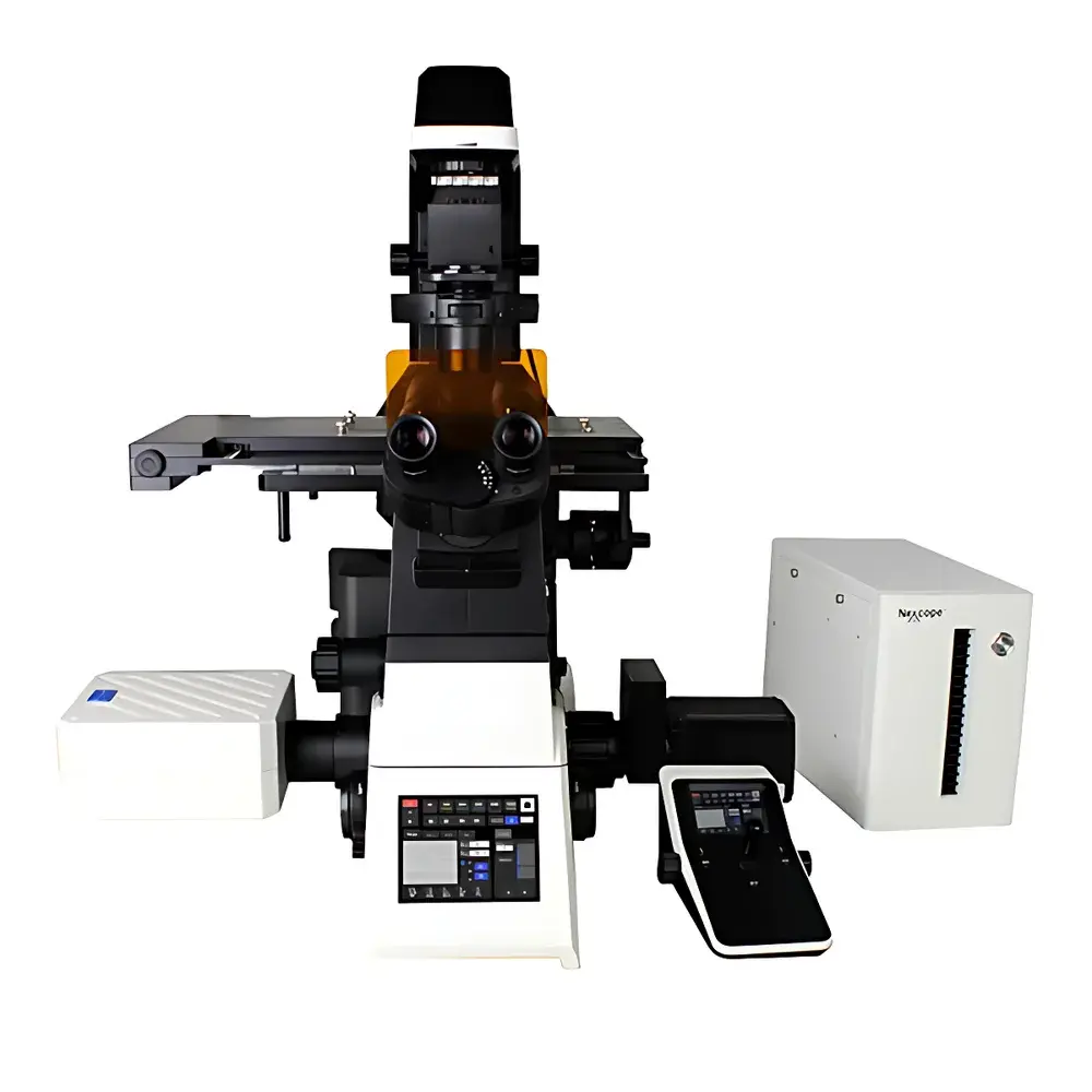
Nexcope NCF1000 Laser Scanning Confocal Microscope
| Brand | Nexcope |
|---|---|
| Origin | Zhejiang, China |
| Model | NCF1000 |
| Instrument Type | Point-Scanning Confocal Microscope |
| Field of View | Φ25 mm (25 mm diameter circular field, square imaging area up to 8192 × 8192 pixels) |
| Lasers | 405 nm, 488 nm, 561 nm, 640 nm |
| Detectors | 4-channel PMT (standard), spectral detector (400–750 nm, 1 nm tuning resolution, up to 4 GaAsP PMTs), transmission PMT detector |
| Scan Modes | X, Y, Z, λ (spectral), and T (time-lapse) — fully combinable |
| Pinhole | Motorized continuous adjustment |
| Objective Lenses | 4×/0.16, 10×/0.45, 20×/0.75, 40×/0.95, 60×/1.42, 100×/1.45 & 100×/1.49 (oil immersion, coverslip-corrected) |
| Microscope Host | NIB1000-AT motorized inverted research-grade platform with NIS60 infinity optical system |
| Illumination | Epifluorescence LED light source (4-band, 6-position motorized filter turret: B/G/U standard |
| Software | Nomis Pro X-C (confocal scanning control, multi-dimensional acquisition, channel presets, hardware-integrated operation, 16-bit image output, real-time deconvolution & basic filtering) |
| XY Stage | Motorized grating-based, 130 mm × 100 mm travel, 0.1 µm positioning accuracy, 0.5 µm repeatability |
| Z Focus | Motorized, 10 mm range, 0.02 µm minimum step, 0.1 µm repeatability |
| Adaptive Focus Stabilization (AFS) | Real-time focal plane maintenance during time-lapse imaging |
| Optional Modules | SR-SIM super-resolution upgrade (2D-SIM lateral resolution ≤85 nm, 3D-SIM axial resolution ≤270 nm |
Overview
The Nexcope NCF1000 Laser Scanning Confocal Microscope is a high-performance, research-grade point-scanning confocal system engineered for quantitative fluorescence imaging in life science laboratories. Built upon the NIB1000-AT motorized inverted microscope platform and integrated with the NIS60 infinity-corrected optical architecture, the NCF1000 delivers diffraction-limited optical sectioning via pinhole-based rejection of out-of-focus light—enabling precise three-dimensional reconstruction of subcellular structures. Its defining feature is the 25 mm field-of-view (FOV) optical design, supporting seamless wide-area imaging without tiling artifacts while maintaining full compatibility with high-magnification, high-numerical-aperture objectives (up to NA 1.49). The system supports concurrent X-Y-Z-λ-T dimensional acquisition, allowing synchronized spatial, spectral, and temporal data capture—essential for dynamic co-localization studies, calcium signaling kinetics, and multicolor FRET assays. All optical paths—including epifluorescence, DIC, phase contrast, and brightfield—are accessible through a modular dual-light-path configuration, ensuring experimental flexibility without mechanical reconfiguration.
Key Features
- 25 mm circular FOV with native 8192 × 8192 pixel scan capability—enabling high-fidelity, single-shot imaging across large specimens (e.g., organoids, tissue explants, micro-patterned substrates)
- Four solid-state lasers (405/488/561/640 nm) with independently controllable intensity and TTL synchronization for multiplexed excitation
- 4-channel PMT detection system with spectral unmixing support; optional GaAsP detectors for enhanced quantum efficiency in the 400–750 nm range
- Motorized, continuously adjustable pinhole (0–3 Airy units) for optimized signal-to-noise ratio versus axial resolution trade-off
- NIB1000-AT host platform featuring motorized 6-position objective turret, 6-position fluorescence filter wheel, and 7-position motorized condenser turret
- Adaptive Focus Stabilization (AFS) system—actively compensates for thermal drift and mechanical creep during extended time-lapse experiments (≥24 h)
- Grating-based XY stage with 130 × 100 mm travel, 0.1 µm closed-loop positioning accuracy, and programmable multi-point acquisition routines
Sample Compatibility & Compliance
The NCF1000 accommodates standard biological sample formats including glass-bottom dishes (35–65 mm), multi-well plates (6–96-well), histological sections (1–50 µm thickness), and live-cell chambers (CO₂/O₂/temperature-controlled via optional SR-Live module). Its optical design complies with ISO 19012-1:2021 (microscope terminology and performance specifications) and supports GLP/GMP-aligned workflows through audit-trail-enabled software logging (Nomis Pro X-C). Data integrity adheres to FDA 21 CFR Part 11 requirements when deployed with electronic signature and role-based access control configurations. All fluorescence filter sets meet ISO/TR 18357:2017 spectral transmission standards for quantitative intensity calibration. The system is compatible with common labeling protocols (e.g., immunofluorescence, live dyes, GFP/RFP fusions) and validated for use with commercial mounting media (e.g., ProLong Diamond, Vectashield).
Software & Data Management
Nomis Pro X-C is a modular, hardware-integrated acquisition and analysis platform supporting multi-dimensional confocal imaging (Z-stacks, time series, spectral lambda stacks, and multi-position tile scans). It provides real-time hardware control—including laser power modulation, PMT gain/voltage, pinhole size, scan speed, and objective selection—via direct driver-level communication. Image data are acquired at native 16-bit depth and stored in open-format TIFF or OME-TIFF containers compliant with Bio-Formats. The software includes built-in background subtraction, flat-field correction, and Gaussian/Median filtering; batch processing pipelines support automated channel alignment, intensity normalization, and Z-projection. Export options include PNG, JPEG2000, and HDF5 for downstream analysis in Fiji/ImageJ, MATLAB, or Python-based tools (e.g., scikit-image, napari). Audit trails record all acquisition parameters, user actions, and timestamped metadata per image file—supporting traceability in regulated environments.
Applications
The NCF1000 serves as a primary imaging platform for cell biology, neuroscience, developmental biology, and pharmaceutical screening. Typical applications include: quantitative co-localization analysis of membrane receptors and cytoskeletal components; 3D reconstruction of neuronal dendritic spines and synaptic puncta; longitudinal tracking of organelle dynamics (mitochondria, lysosomes, Golgi) under pharmacological perturbation; high-content imaging of stem cell differentiation in 3D hydrogels; and spectral unmixing of autofluorescence interference in tissue sections. Its wide FOV and motorized precision make it suitable for high-throughput phenotypic screening (e.g., siRNA knockdown libraries), while its compatibility with SR-SIM modules extends utility into nanoscale structural analysis—such as nuclear pore complex organization or clathrin-coated pit assembly—without requiring specialized training in STED or PALM methodologies.
FAQ
What is the maximum usable field of view for confocal imaging on the NCF1000?
The system supports a true 25 mm diameter field of view with full optical fidelity; the maximum native scan area is 8192 × 8192 pixels at 1:1 pixel-to-detector mapping, scalable across magnifications via digital zoom without interpolation.
Does the NCF1000 support spectral unmixing and linear unmixing algorithms?
Yes—Nomis Pro X-C includes real-time spectral unmixing using reference spectra libraries and supports custom emission profile import for linear unmixing of up to four fluorophores with overlapping spectra.
Can the system be upgraded to super-resolution imaging post-purchase?
Yes—the SR-SIM module is field-installable and fully integrated into Nomis Pro X-C; no hardware modification to the base confocal scan head is required.
Is the NIB1000-AT microscope body compatible with third-party accessories (e.g., micromanipulators, perfusion systems)?
Yes—the NIB1000-AT features standardized dovetail rails, C-mount and M27 threaded ports, and TTL/RS-232 interfaces for synchronization with external devices such as patch-clamp amplifiers and piezoelectric Z-drives.
What validation documentation is provided for regulatory compliance?
Nexcope supplies IQ/OQ documentation templates, laser power calibration certificates (traceable to NIST standards), and software validation packages supporting 21 CFR Part 11 implementation—including electronic signature configuration guides and change control logs.


