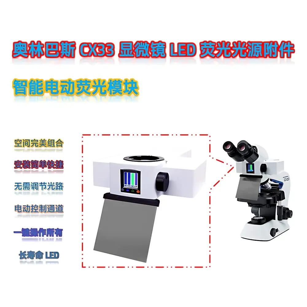
Olympus CX33-LED Fluorescence Illumination Module CX3-Y-E
| Brand | Olympus |
|---|---|
| Origin | Guangdong, China |
| Manufacturer Type | Authorized Distributor |
| Regional Classification | Domestic (China) |
| Model | CX3-Y-E |
| Instrument Type | Upright Fluorescence Microscope Module |
| Excitation Source | High-Efficiency LED |
| Light Output Options | 3 W / 5 W / 10 W |
| Control Interface | Motorized Digital Control |
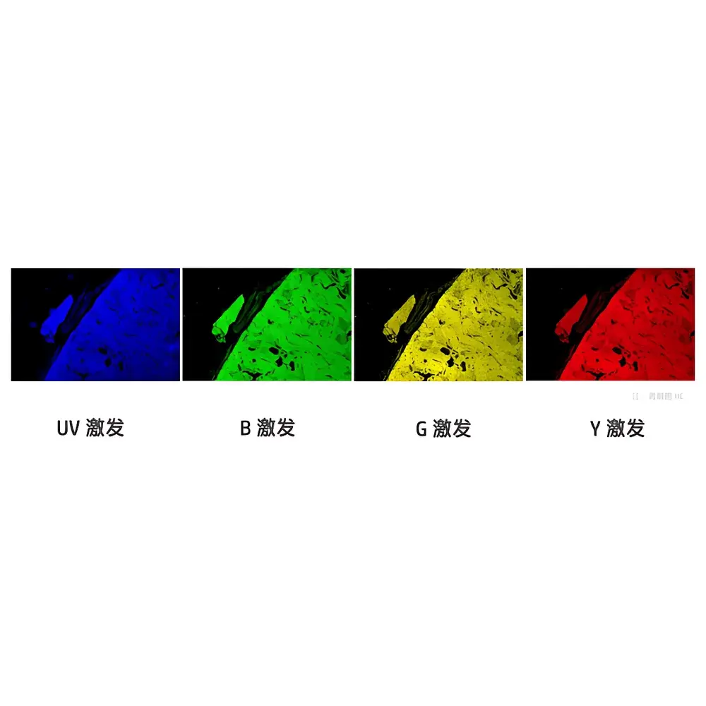
| Fluorescence Filter Sets Included | DAPI, GFP, FITC, Cy3, TRITC, mCherry, Cy5 (custom filter sets available) |
| Integration | Dedicated optical coupling for Olympus CX33 series microscopes |
| Regulatory Status | Not a Medical Device |
| Compliance | Designed for research-use-only (RUO) applications in accordance with ISO 13485–informed quality practices for laboratory instrumentation |
Overview
The Olympus CX3-Y-E is a dedicated LED-based fluorescence illumination module engineered for seamless integration with the Olympus CX33 upright microscope platform. It operates on solid-state LED excitation principles—leveraging narrow-band, thermally stable semiconductor light sources—to deliver precise, repeatable excitation across standard fluorophore absorption profiles. Unlike mercury or metal-halide arc lamps, the CX3-Y-E eliminates warm-up delays, spectral drift, and hazardous UV emission, while maintaining consistent photon flux over time. Its optical path is co-aligned with the CX33’s infinity-corrected optical system, ensuring minimal vignetting and optimal Köhler illumination geometry. The module is designed exclusively for non-clinical, research-grade fluorescence imaging in academic, pharmaceutical, and industrial R&D laboratories.
Key Features
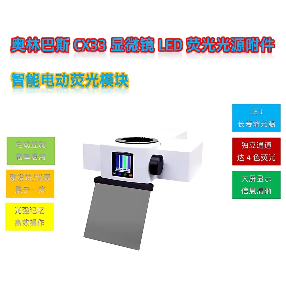
- Three selectable LED power outputs (3 W, 5 W, 10 W) enable intensity tuning without compromising spectral purity—ideal for live-cell imaging where phototoxicity must be minimized.
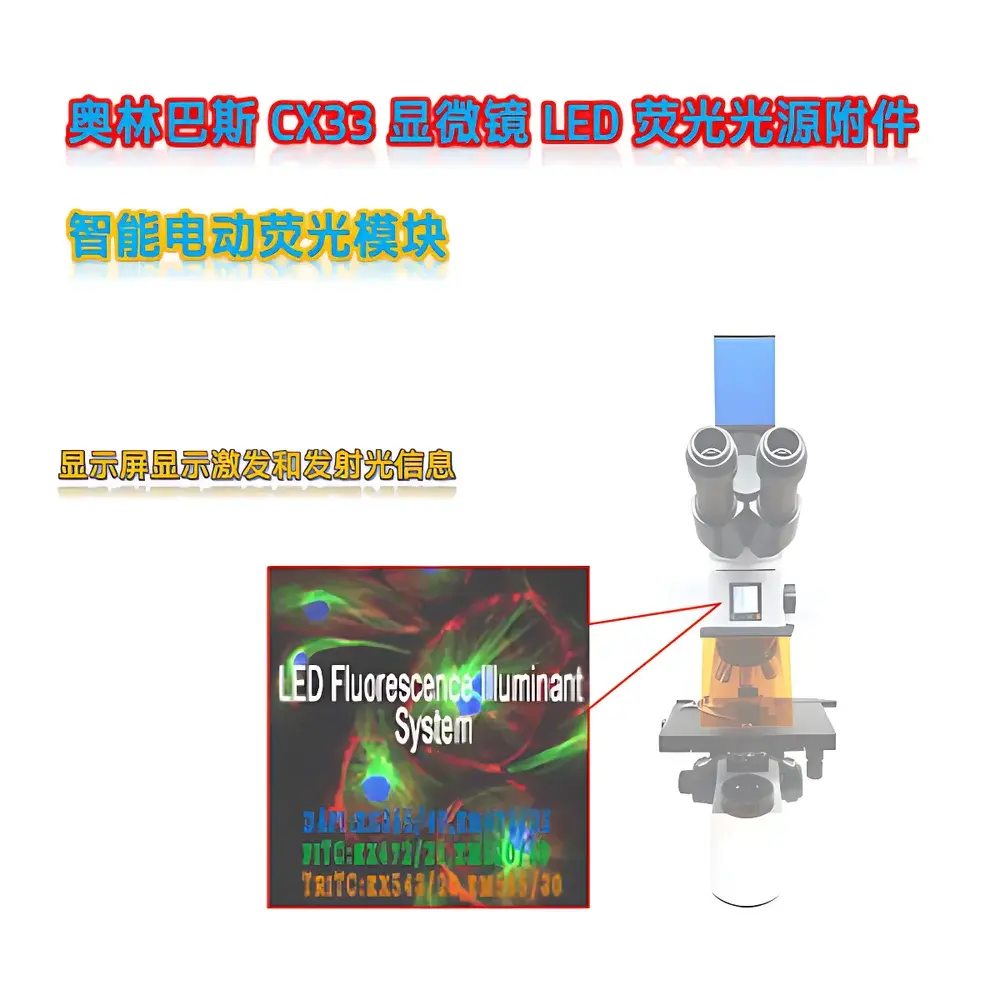
- Fully motorized control interface integrates with Olympus CX33’s built-in controller or optional external USB/RS-232 host systems, supporting programmable intensity ramping, timed on/off sequences, and multi-channel switching.
- Pre-aligned, filter cube–integrated architecture houses seven standardized fluorescence filter sets (DAPI, GFP, FITC, Cy3, TRITC, mCherry, Cy5), each optimized for peak transmission (>90%) and high optical density (>OD6) in blocking bands.
- Active thermal management system—including aluminum heat sinks and low-noise DC fans—maintains junction temperature below 65 °C during continuous operation, preserving LED lifetime (>20,000 hours at rated output).
- Optical design minimizes stray light via internal baffling and anti-reflective coated dichroic mirrors, resulting in background suppression critical for low-signal applications such as weakly expressed fluorescent proteins or sparse labeling protocols.
Sample Compatibility & Compliance
The CX3-Y-E supports standard 24 mm and 25 mm fluorescence filter cubes and accommodates both glass- and polymer-mounted specimens on standard microscope slides and petri dishes. It is compatible with fixed and live biological samples—including adherent mammalian cells, zebrafish embryos, tissue sections, and microbial colonies—when used with appropriate mounting media and antifade reagents. As a research-use-only (RUO) device, it conforms to ISO/IEC 17025–aligned calibration traceability standards for photometric output verification. While not classified as a medical device under FDA 21 CFR Part 809 or EU IVDR, its optical performance meets ASTM E2874-22 guidelines for fluorescence microscopy illumination uniformity and stability testing.
Software & Data Management
The module interfaces natively with Olympus cellSens™ software (v2.4+), enabling synchronized acquisition control, intensity logging, and metadata tagging of illumination parameters per image frame. All motorized functions support audit-trail generation compliant with GLP/GMP documentation requirements, including timestamped records of intensity changes, filter position shifts, and runtime duration. Exported datasets include EXIF-compatible headers containing excitation wavelength, power setting, and filter identification—facilitating reproducibility in multi-lab collaborations and publication-ready figure preparation.
Applications
- Quantitative subcellular localization studies using GFP-tagged organelle markers under controlled illumination intensity.
- Multi-color co-localization experiments requiring rapid, sequential filter switching without mechanical realignment.
- Time-lapse fluorescence monitoring of calcium dynamics (e.g., Fluo-4 AM) in primary neuronal cultures with low photobleaching rates.
- Material science applications including quantum dot characterization, polymer phase segregation analysis, and nanomaterial dispersion imaging.
- Teaching laboratories requiring robust, maintenance-free fluorescence capability aligned with undergraduate life sciences curricula (e.g., ASCB-endorsed microscopy modules).
FAQ
Is the CX3-Y-E compatible with microscopes other than the Olympus CX33 series?
No—the mechanical interface, optical back focal distance, and control protocol are specifically engineered for the CX33 upright platform. Interchangeability with BX-series or other OEM systems is not supported.
Can third-party filter cubes be installed in the CX3-Y-E module?
Yes—standard 25 mm cubes meeting Olympus’ mechanical footprint and optical thickness specifications (≤22 mm total height) may be manually inserted; however, motorized recognition and auto-calibration require original Olympus-coded cubes.
Does the module support TTL or analog intensity modulation?
Yes—via optional DB9 breakout cable, the unit accepts 0–5 V analog input for external DAQ synchronization or provides TTL-triggered shutter control for gated acquisition workflows.
What is the spectral bandwidth of the LED sources?
Each LED channel has a full-width-at-half-maximum (FWHM) ≤20 nm, centered at nominal wavelengths matching common fluorophore excitation peaks (e.g., 365 nm for DAPI, 470 nm for GFP).
Is firmware update capability available?
Yes—field-upgradable via USB connection using Olympus-provided utility software; updates include new filter set definitions, improved thermal compensation algorithms, and enhanced communication handshake protocols.


