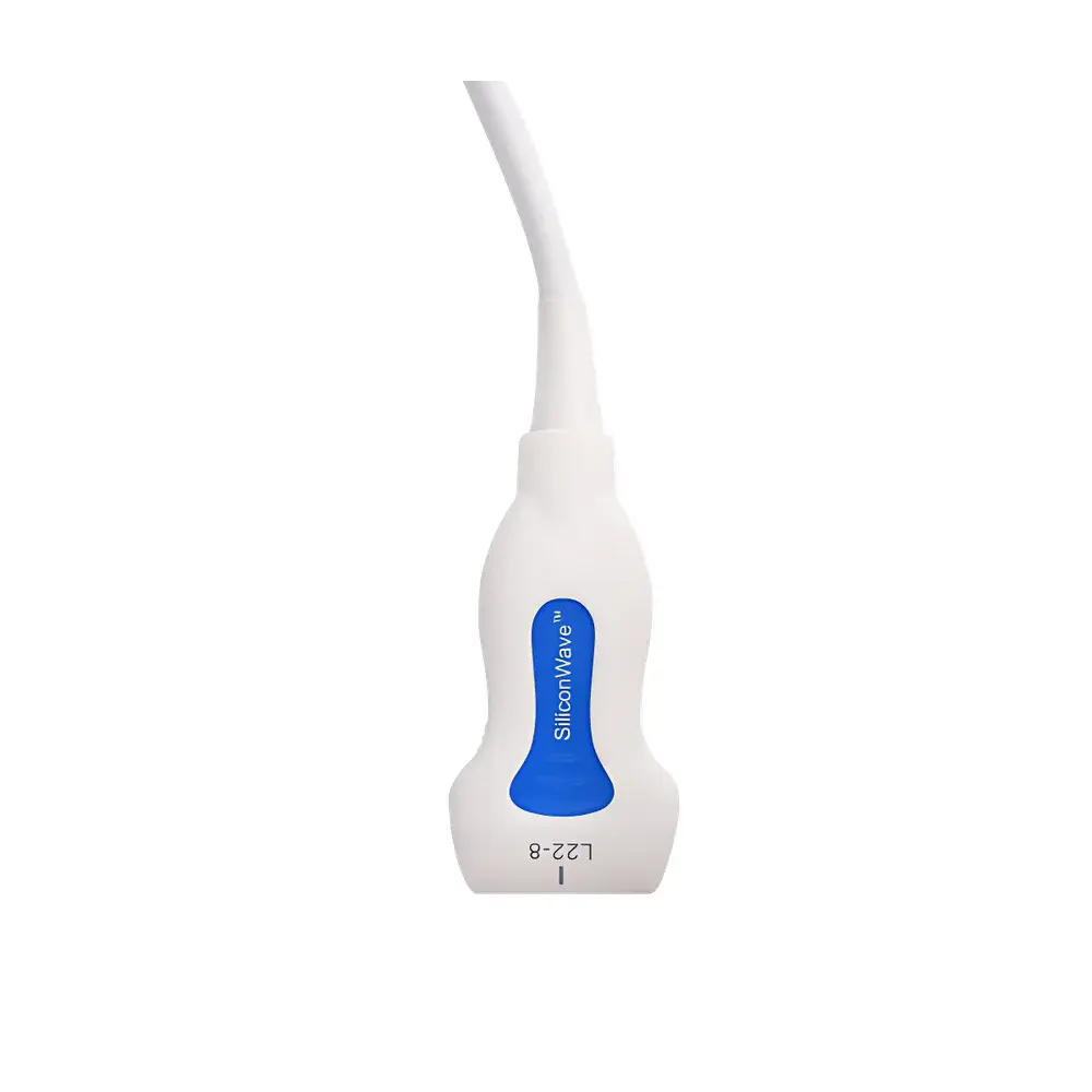
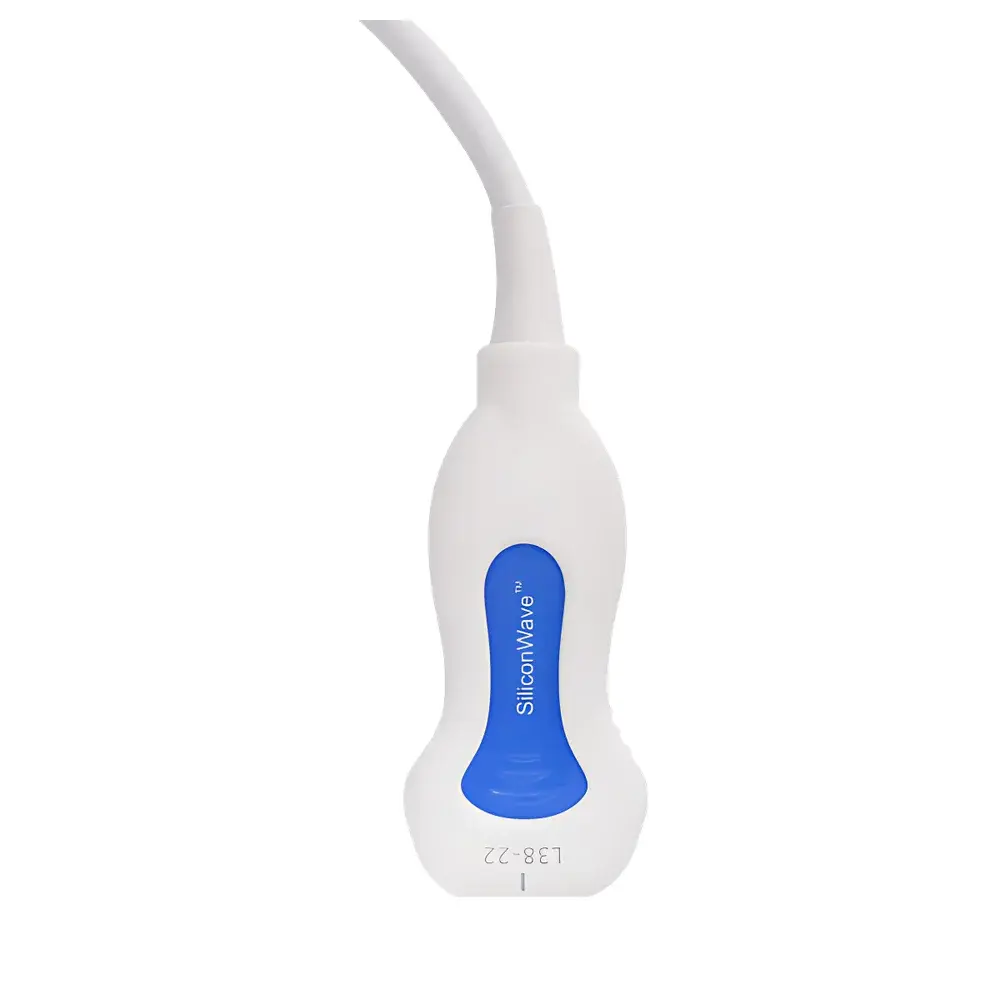
Paragon XHD Ultra-High-Frequency Preclinical Ultrasound Imaging System
| Origin | Jiangsu, China |
|---|---|
| Manufacturer Type | Distributor |
| Origin Category | Domestic |
| Model | Paragon XHD |
| Price Range | USD 0–140,000 (FOB) |
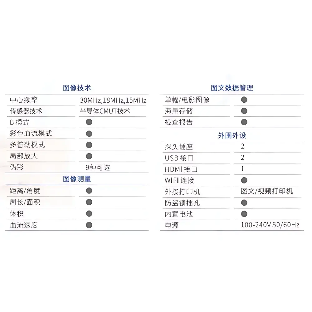
| Spatial Resolution | 80–100 µm |
| Temporal Resolution | 30 fps |
| Gray Scale | 256 levels |
| Time-Gain Compensation (TGC) | 8 adjustable segments |
| Measurement Functions | Distance, Circumference, Area, Volume, Depth, Angle |
| Dynamic Range | 30–150 dB |
| Display Modes | B-mode + Color Doppler + Pulsed-Wave Doppler (B+C+PW) |
| Maximum Penetration Depth | 50 mm |
Overview
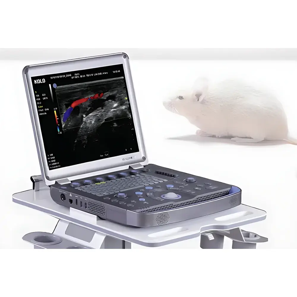
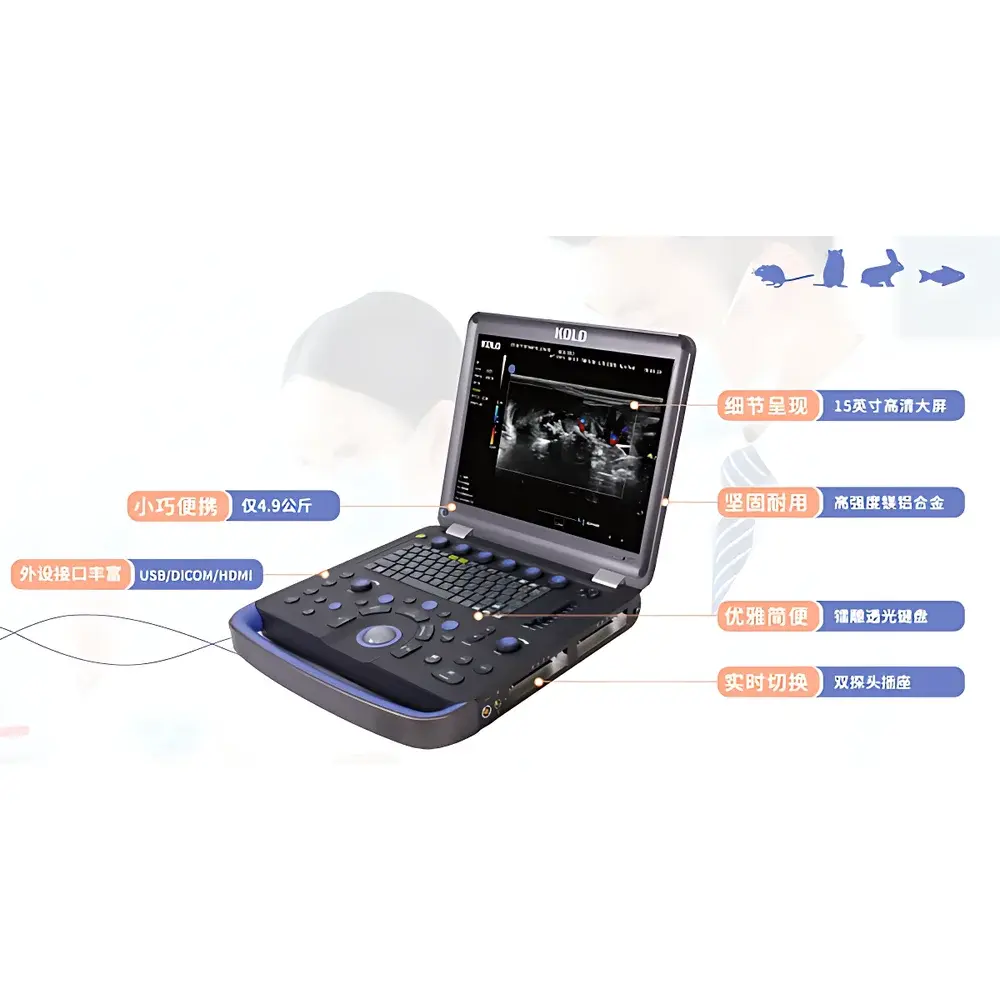
The Paragon XHD is a state-of-the-art ultra-high-frequency (UHF) preclinical ultrasound imaging system engineered specifically for high-resolution, non-invasive in vivo visualization of small animal anatomy and hemodynamics. Operating at center frequencies exceeding 40 MHz—enabled by proprietary Capacitive Micromachined Ultrasonic Transducer (CMUT) semiconductor technology—the system delivers microstructural detail unattainable with conventional piezoelectric transducers. CMUT architecture provides superior bandwidth, sensitivity, and thermal stability, supporting consistent acquisition of both structural (B-mode) and functional (Color and PW Doppler) data across longitudinal studies. Designed for rigorous laboratory environments, the Paragon XHD meets the demanding requirements of translational research where quantitative reproducibility, spatial fidelity, and temporal consistency are critical to hypothesis validation.
Key Features
- Microscale Spatial Resolution: Achieves 80–100 µm axial and lateral resolution at near-surface depths, enabling visualization of capillary networks, tumor microvasculature, embryonic structures, and murine cardiac wall motion with sub-millimeter anatomical fidelity.
- Real-Time Multimodal Imaging: Simultaneous B-mode, Color Doppler, and Pulsed-Wave Doppler display (B+C+PW) supports concurrent morphological assessment and quantitative flow analysis—including peak systolic velocity, resistive index, and pulsatility index—without frame interpolation or mode switching latency.
- Adaptive Signal Processing: Eight-segment Time-Gain Compensation (TGC) allows precise depth-dependent amplification control, while a 120 dB dynamic range (30–150 dB) ensures optimal contrast differentiation across heterogeneous tissue interfaces—from superficial dermis to deep renal parenchyma within the 50 mm penetration limit.
- Quantitative Measurement Suite: Integrated on-screen calipers support ISO/IEC 17025-aligned measurement protocols for distance, circumference, area, volume, depth, and angle—traceable to NIST-calibrated phantoms and compliant with GLP documentation standards.
- Thermally Stable CMUT Architecture: Silicon-based transducer arrays eliminate piezoelectric hysteresis and exhibit minimal frequency drift under continuous operation, ensuring measurement repeatability across multi-hour imaging sessions and multi-day longitudinal experiments.
Sample Compatibility & Compliance
The Paragon XHD is validated for use with murine (mouse, rat), lagomorph (rabbit), and zebrafish models. Its compact footprint and ergonomic transducer holder accommodate stereotactic positioning systems and temperature-controlled imaging chambers (28–37 °C). The system complies with IEC 62359:2015 (ultrasound equipment safety), IEC 61223-3-5:2019 (acceptance testing for medical imaging devices), and supports audit-ready data export conforming to DICOM Supplement 157 (Ultrasound Image Storage). All measurement metadata—including TGC settings, gain, depth, frame rate, and Doppler angle—are embedded in DICOM headers for regulatory traceability per FDA 21 CFR Part 11 and EU MDR Annex II requirements.
Software & Data Management
Built on a real-time Linux-based acquisition platform, the Paragon XHD’s proprietary software (v4.2+) provides DICOM-compliant image archiving, encrypted study-level database management, and batch-processing pipelines for volumetric reconstruction and Doppler quantification. Raw RF data export (in HDF5 format) enables third-party algorithm integration (e.g., MATLAB, Python-based elastography or flow modeling). Audit logs record user login, parameter modification timestamps, and image annotation history—fully compatible with GLP/GMP electronic record retention policies. Software updates follow ISO 13485 design control procedures and include version-signed release notes for validation documentation.
Applications
- Oncology: Longitudinal monitoring of subcutaneous and orthotopic tumor angiogenesis, necrosis, and treatment response via 3D vascular density mapping and perfusion kinetics.
- Cardiovascular Research: High-frame-rate assessment of murine left ventricular ejection fraction, diastolic filling dynamics, and valvular regurgitation using ECG-gated cine loops.
- Developmental Biology: In utero imaging of embryonic heart tube formation, neural tube closure, and placental vascular remodeling in timed-pregnant mice.
- Pharmacokinetics & Toxicology: Real-time visualization of nanoparticle biodistribution and organ-specific accumulation following IV administration.
- Urology & Reproductive Medicine: Quantitative evaluation of testicular blood flow, ovarian follicle maturation, and bladder wall thickness changes in disease models.
FAQ
What is the maximum imaging depth achievable with the Paragon XHD system?
The system achieves a maximum penetration depth of 50 mm in soft tissue-equivalent phantoms; effective depth varies with frequency selection and acoustic coupling—optimal resolution is maintained within the first 15 mm for 60 MHz imaging.
Does the Paragon XHD support 3D/4D reconstruction?
Yes—motorized linear and rotational transducer mounts (optional accessories) enable automated volumetric sweeps; reconstructed datasets are exportable as NIfTI or STL for downstream segmentation and finite-element modeling.
Is the system compatible with existing preclinical infrastructure such as anesthesia systems or physiological monitors?
The Paragon XHD features analog and digital I/O ports (TTL, RS-232, USB-C) for hardware synchronization with gas anesthesia controllers, ECG amplifiers, and respiration gating modules.
How is calibration maintained across multiple users and long-term studies?
The system includes daily self-calibration routines using built-in reference phantoms; all calibration events are timestamped and logged in the audit trail, supporting ISO/IEC 17025 method validation requirements.
Can raw RF data be exported for custom signal processing?
Yes—HDF5-formatted RF data streams (including IQ samples, beamformed channel data, and metadata) are accessible via secure API, enabling implementation of advanced beamforming, clutter filtering, or machine learning–based tissue classification algorithms.


")