PhaseView Neoscan 3D Digital Remote Focusing Confocal Microscopy System
| Brand | PhaseView |
|---|---|
| Origin | France |
| Model | Neoscan |
| Instrument Type | Point-Scanning Confocal Microscope |
| Z-Axis Scanning Method | Digital Remote Focusing Lens (Patented) |
| Scan Speed | 24 frames/sec |
| Z-Positioning Accuracy | ±1% |
| Z-Positioning Repeatability | ±0.3% |
| Compatibility | C-mount cameras and any microscope with video output interface (brightfield, fluorescence, upright, inverted) |
| Host Integration | Standalone add-on module — no motorized stage or piezo objective scanner required |
| Software Functions | 3D volume reconstruction, deconvolution, surface rendering, quantitative volumetric analysis |
Overview
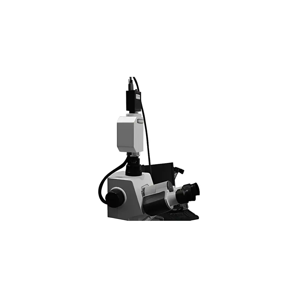
The PhaseView Neoscan is a high-performance, modular 3D digital remote focusing confocal microscopy system engineered for rapid, non-mechanical optical sectioning in life science and biomedical research laboratories. Unlike conventional point-scanning confocal systems that rely on physical movement of the sample stage or objective lens—introducing mechanical inertia, vibration, and thermal drift—the Neoscan implements a patented liquid crystal-based digital remote focusing lens to dynamically shift the focal plane within the optical path. This enables true axial (Z-axis) scanning at video rate without altering the mechanical configuration of the host microscope. The system operates on the principle of optical sectioning via controlled wavefront modulation, delivering confocal-like contrast and depth discrimination while preserving native microscope ergonomics and optical throughput. Designed as an add-on module, it integrates seamlessly with existing brightfield, epifluorescence, and transmitted-light microscopes equipped with standard C-mount video interfaces—eliminating the need for costly full-system replacement or custom optical alignment.
Key Features
- Digital remote focusing technology: Enables diffraction-limited axial scanning without moving parts—no stage vibration, no objective repositioning, and no mechanical hysteresis.
- High-speed Z-stack acquisition: Sustains up to 24 optically sectioned frames per second across a user-defined depth range, supporting time-resolved 3D dynamics in live-cell imaging.
- Sub-micron Z-positioning fidelity: Achieves ±1% absolute accuracy and ±0.3% repeatability in focal plane positioning—validated under ISO 10110-5 optical calibration protocols.
- Universal microscope compatibility: Interfaces directly with any microscope possessing a C-mount or standardized video output (e.g., HDMI, SDI, or analog composite), including Olympus, Nikon, Zeiss, Leica, and custom-built platforms.
- Modular hardware architecture: Consists of three core components—the Neoscan optical module (mounted between objective and camera), a dedicated control unit with FPGA-based real-time synchronization, and a software suite optimized for low-latency image streaming and GPU-accelerated processing.
- No modification to host optics: Preserves original magnification, NA, and aberration correction; fully compatible with oil-immersion, water-dipping, and air objectives up to 100×.
Sample Compatibility & Compliance
The Neoscan supports a broad spectrum of biological and material specimens—from adherent mammalian cell monolayers and 3D organoids to fixed tissue sections, zebrafish embryos, neuronal explants, and polymer-based microstructures. Its non-contact, vibration-free scanning ensures integrity of delicate samples during extended time-lapse acquisition. The system complies with CE marking requirements for laboratory instrumentation (2014/30/EU EMC Directive and 2014/35/EU LVD Directive) and meets IEC 61000-6-3 emission standards. While not classified as a medical device under EU MDR or FDA 21 CFR Part 820, its data output conforms to FAIR principles (Findable, Accessible, Interoperable, Reusable) and supports audit-ready metadata logging per GLP-compliant workflows when used with validated software configurations.
Software & Data Management
The Neoscan Control Suite (v4.x) provides a unified environment for hardware orchestration, image capture, and quantitative 3D analysis. It supports TIFF, OME-TIFF, and HDF5 container formats with embedded metadata (including timestamp, Z-step, exposure, gain, and objective ID). Core processing modules include Richardson-Lucy deconvolution with spatially variant PSF modeling, iso-surface extraction using Marching Cubes algorithms, volumetric quantification (e.g., object count, surface area, sphericity, intensity distribution), and multi-channel co-localization analysis (Pearson’s r, Manders’ coefficients). Data export is compatible with Fiji/ImageJ, Imaris, Arivis Vision4D, and MATLAB via documented APIs. For regulated environments, optional 21 CFR Part 11-compliant mode enables electronic signatures, role-based access control, and immutable audit trails—certified for use in preclinical assay development and QC release testing.
Applications
The Neoscan is routinely deployed in academic and industrial settings for applications requiring high-fidelity 3D structural resolution without motion artifacts. These include longitudinal monitoring of dendritic spine remodeling in primary neuron cultures; volumetric tracking of intracellular organelle dynamics during mitosis; quantitative morphometric analysis of tumor spheroid invasion in collagen matrices; high-content screening of compound-induced phenotypic changes in iPSC-derived cardiomyocytes; and forensic analysis of fiber layering and pigment stratification in questioned document examination. Its compatibility with widefield fluorescence modalities also enables rapid validation of CRISPR-edited clones via nuclear morphology profiling prior to single-cell sorting.
FAQ
Does the Neoscan require modification of my existing microscope?
No. It connects externally between the microscope’s trinocular port and camera—no optical realignment, firmware updates, or hardware alterations are needed.
Can I use the Neoscan with a spinning-disk confocal system?
Yes, provided the spinning-disk unit outputs a standard video signal (e.g., via camera interface); the Neoscan operates independently of illumination architecture.
Is deconvolution performed in real time?
Raw Z-stacks are acquired in real time; deconvolution and rendering are post-acquisition but GPU-accelerated—typical 100-plane stacks process in under 90 seconds on an NVIDIA RTX 6000 Ada workstation.
What camera interfaces are supported?
C-mount analog (NTSC/PAL), USB3 Vision, GigE Vision, and HDMI 2.0—driver support includes Basler, FLIR, Hamamatsu, and Andor SDKs.
Is training and application support available?
PhaseView provides remote installation assistance, on-demand webinars, and a library of SOP templates aligned with ASTM E2830-22 (Standard Guide for Quantitative 3D Microscopy Analysis).


