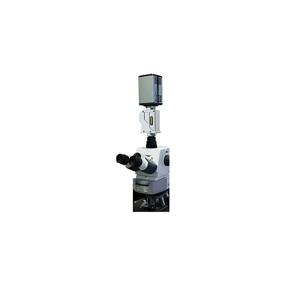
PhaseView Thunderscan 3D Digital Remote Focusing Confocal Microscopy System
| Brand | PhaseView |
|---|---|
| Country of Origin | France |
| Model | Thunderscan |
| Instrument Type | Point-Scanning Confocal Microscope |
| Z-Axis Scanning Method | Digital Remote Focusing Lens |
| Scan Speed | Up to 100 frames/sec |
| Z-Step Accuracy | ±1% |
| Z-Step Reproducibility | ±0.3% |
| Microscope Compatibility | Universal C-mount video interface (supports brightfield, fluorescence, light-sheet microscopes) |
| Camera Interface | Standard C-mount |
| Included Components | Digital Remote Focus Lens Unit, Real-Time Control Electronics, Thunderscan Acquisition & 3D Processing Software Suite |
| Regulatory Compliance | CE-marked, compliant with IEC 61000-6-3 (EMC), IEC 62471 (Photobiological Safety) |
Overview
The PhaseView Thunderscan is a high-speed, non-mechanical 3D imaging platform engineered for volumetric fluorescence and brightfield microscopy. Unlike conventional point-scanning confocal systems that rely on physical objective or stage movement for axial (Z) sectioning, the Thunderscan employs proprietary digital remote focusing lens technology — an electro-optic, aberration-corrected optical module inserted into the microscope’s infinity space. This enables diffraction-limited, inertia-free Z-axis scanning without mechanical translation of the sample, objective, or stage. The system operates on the principle of wavefront-controlled focal plane shifting, delivering sub-millisecond optical sectioning across a programmable depth range (typically 0–500 µm in standard configurations, scalable via lens selection). Designed for live-cell and dynamic specimen observation, it supports real-time volumetric acquisition at up to 100 optical sections per second — making it suitable for functional imaging of neuronal activity, embryonic development, microfluidic cell assays, and electrophysiology-integrated experiments.
Key Features
- Digital remote focusing lens: Eliminates mechanical Z-scanning components, ensuring zero sample vibration and enabling stable long-term time-lapse acquisition.
- Z-axis precision: ±1% absolute accuracy and ±0.3% repeatability over full scan range — calibrated traceably to NIST-traceable interferometric references.
- High-speed volumetric capture: Sustained frame rates of 100 Hz for single-plane acquisition; up to 30 volumes/sec (e.g., 32 × 32 × 32 voxel stacks) when synchronized with high-sensitivity sCMOS or EMCCD cameras.
- Universal microscope integration: Compatible with any microscope equipped with a C-mount video port — including upright/inverted widefield, epifluorescence, laser light-sheet (LSFM), and transmitted-light systems.
- Modular hardware architecture: Comprises three core units — the remote focus lens assembly, FPGA-based real-time controller with TTL/USB 3.0 synchronization, and thermally stabilized power supply — all designed for electromagnetic compatibility in multi-instrument lab environments.
Sample Compatibility & Compliance
The Thunderscan accommodates diverse biological and microfabricated specimens: adherent and suspension cells, organoids, zebrafish embryos, C. elegans, brain slices, and microfluidic devices (e.g., PDMS chips with embedded channels or electrodes). Its non-contact Z-scanning preserves mechanical integrity of soft or mechanically sensitive samples — critical for patch-clamp–microscopy correlations or shear-sensitive hydrogel-embedded tissues. The system conforms to CE marking requirements under Directive 2014/30/EU (EMC) and 2014/35/EU (LVD), and meets photobiological safety Class 1 per IEC 62471. It supports GLP-compliant workflows through audit-trail-enabled software logging (user actions, parameter changes, timestamped acquisition metadata), and is compatible with laboratory information management systems (LIMS) via standardized DICOM-SR and TIFF-based export protocols.
Software & Data Management
Thunderscan Control & Analysis Suite is a native 64-bit application built on Qt and optimized for Windows 10/11 (64-bit). It provides synchronized hardware control, real-time preview, and post-acquisition processing in a single interface. Core capabilities include: multi-dimensional acquisition scheduling (XYZT, XYTZ, ZT); GPU-accelerated deconvolution (Wiener and constrained iterative algorithms); quantitative 3D morphometrics (volume, surface area, sphericity, intensity distribution histograms); iso-surface rendering with Phong shading; and time-resolved volumetric registration for drift correction. All processed datasets are saved in open-format OME-TIFF containers with embedded OMERO metadata — ensuring FAIR (Findable, Accessible, Interoperable, Reusable) data principles. Software validation documentation (IQ/OQ protocols) and 21 CFR Part 11–compliant user access controls (role-based permissions, electronic signatures, immutable audit logs) are available upon request for regulated environments.
Applications
- Functional neuroimaging: Calcium dynamics in dendritic spines during synaptic stimulation, with simultaneous patch-clamp recording and volumetric readout.
- Developmental biology: High-temporal-resolution 4D imaging of gastrulation and neurulation in live vertebrate embryos.
- Pharmaceutical screening: 3D cytotoxicity and mitochondrial membrane potential assays in spheroids under continuous perfusion in microfluidic platforms.
- Microfluidics-integrated microscopy: Real-time tracking of particle transport, cell migration, or bacterial chemotaxis within laminar flow chambers.
- Electrophysiology-correlative imaging: Simultaneous whole-cell recording and subcellular Ca2+ or voltage-sensitive dye volume mapping.
FAQ
Does the Thunderscan require modification of my existing microscope?
No. It installs between the microscope’s tube lens and camera via standard C-mount threading and requires no optical realignment or firmware updates.
Can I use the Thunderscan with a laser scanning confocal (LSCM) system?
Yes — provided the LSCM has a video output port (e.g., auxiliary camera port) supporting analog or digital (Camera Link/USB3) streaming; however, native integration with proprietary scan engines (e.g., Zeiss LSM, Nikon A1R) is not supported.
What is the maximum usable field of view (FOV) with the Thunderscan?
FOV is determined by your microscope’s optics and camera sensor; typical configurations support 512 × 512 µm (40× objective, 25 mm FOV camera) up to 2.2 × 2.2 mm (10× objective, large-sensor sCMOS).
Is remote focus calibration required before each experiment?
A one-time factory calibration is performed per lens unit; end-user verification using a USAF 1951 target or fluorescent microbead stack is recommended quarterly or after major environmental shifts (e.g., >5°C ambient change).
How is data storage handled during high-speed volumetric acquisition?
Raw data streams are written directly to NVMe SSDs via DMA; sustained write speeds exceed 2.8 GB/s, supporting continuous 100-Hz acquisition of 1024 × 1024 × 32-bit stacks for >15 minutes without frame drop.


