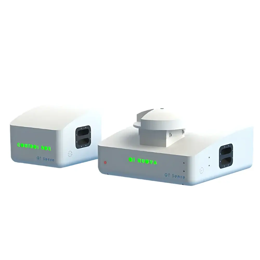
Quant Nuova Quantum Radical Detection System for Biomedical Oxidative Stress Research
| Brand | 1124123/13213112 |
|---|---|
| Origin | Netherlands |
| Manufacturer Type | Authorized Distributor |
| Origin Category | Imported |
| Model | Quant Nuova |
| Pricing | Upon Request |
Overview
The Quant Nuova Quantum Radical Detection System is a research-grade quantum sensing platform engineered for label-free, non-invasive, nanoscale detection and spatiotemporal mapping of reactive oxygen species (ROS) and other paramagnetic free radicals in living biological systems. Unlike conventional biochemical assays (e.g., DCFH-DA fluorescence or ESR spin trapping), this system leverages nitrogen-vacancy (NV) center physics in fluorescent nanodiamonds (FNDs) coupled with optically detected magnetic resonance (ODMR) and confocal microscopy. It operates on the principle that electron spin states of NV centers are exquisitely sensitive to local magnetic field fluctuations induced by nearby unpaired electrons—enabling quantitative, subcellular-resolution radical detection at nanomolar concentrations without perturbing native redox homeostasis. Designed for rigorous biomedical investigation, the system bridges quantum metrology and translational cell biology, supporting mechanistic studies of oxidative stress in aging, neurodegeneration, infertility, infection response, and metabolic dysfunction.
Key Features
- Integrated diamond magnetometer and confocal laser scanning microscope for simultaneous optical imaging and magnetic resonance readout
- Nanodiamond-based quantum sensors with surface-functionalized FNDs (5–30 nm diameter) for intracellular delivery and organelle-targeted ROS monitoring
- ODMR spectroscopy with <10 nT magnetic field sensitivity and <50 ms temporal resolution per voxel
- Sub-diffraction spatial resolution (~250 nm lateral, ~700 nm axial) enabled by stimulated emission depletion (STED)-compatible optical architecture
- Real-time relaxation time (T1, T2*) mapping across live cells, tissues, or 3D organoids under physiological conditions
- Modular design supporting multi-modal correlative workflows: fluorescence lifetime imaging (FLIM), calcium imaging, and phase-contrast tracking
Sample Compatibility & Compliance
The Quant Nuova system is validated for use with primary human cells (e.g., endothelial cells, granulosa cells, spermatozoa), yeast (S. cerevisiae), bacterial cultures (E. coli, S. aureus), and ex vivo tissue sections. Sample preparation adheres to ISO 13485-aligned handling protocols for biospecimen integrity. Data acquisition complies with GLP principles for preclinical research; raw ODMR spectra and relaxation maps are timestamped, user-annotated, and stored with full audit trail metadata. While not a CE-marked IVD device, the platform supports method development aligned with FDA Guidance for Industry on Bioanalytical Method Validation (2018) and ISO/IEC 17025 requirements for measurement uncertainty estimation.
Software & Data Management
Control and analysis are performed via QuantStudio™ v3.2 software—a Python-based suite with modular GUI modules for pulse sequence programming (T1/T2* mapping, Rabi oscillation, dynamical decoupling), automated cell segmentation, and multivariate spectral deconvolution. All datasets conform to MIAME-compliant HDF5 container format with embedded ontologies (OBO Foundry: ROS, cellular component, disease). The software includes built-in machine learning pipelines (XGBoost, U-Net) trained on benchmarked ROS perturbation datasets for predictive classification of oxidative phenotypes. Audit logs meet 21 CFR Part 11 requirements for electronic records and signatures when deployed in regulated environments.
Applications
- Quantitative assessment of mitochondrial ROS burst kinetics in human spermatozoa during cryopreservation
- Mapping spatial heterogeneity of superoxide production in TNF-α-stimulated endothelial monolayers
- Monitoring real-time antioxidant efficacy of polyphenol compounds in primary ovarian granulosa cells
- Correlating NV-center T1 shortening with SARS-CoV-2 spike protein-induced membrane lipid peroxidation in lung organoids
- Resolving subcellular ROS gradients in dendritic spines of Huntington’s disease patient-derived neurons
- High-throughput screening of polymer nanoparticle-induced oxidative burden in macrophage phagolysosomes
- Metabolic flux analysis via redox-coupled NAD+/NADH dynamics inferred from NV spin relaxation rates in yeast
FAQ
Is the Quant Nuova system compatible with standard cell culture incubators?
Yes—the optical head is designed for integration with stage-top environmental chambers (37°C, 5% CO2, humidity control) and features vibration-isolated mounting for long-term time-lapse experiments.
Can it distinguish between different ROS species (e.g., •OH vs. O2•−)?
Not directly via spectral fingerprinting; however, differential relaxation responses under selective pharmacological inhibition (e.g., SOD mimetics, catalase inhibitors) combined with compartment-specific FND targeting enable indirect speciation with high biological context fidelity.
What sample preparation is required beyond standard cell plating?
Cells are incubated with biotinylated or antibody-conjugated FNDs (1–2 hr, 37°C); no fixation, permeabilization, or exogenous dyes are needed. Full protocol documentation—including endotoxin testing certificates for FND batches—is provided.
Does the system support automated multi-well plate scanning?
Yes—integrated motorized XYZ stage and adaptive focus lock enable unattended acquisition across 96- and 384-well formats, with ROI-based triggering for phenotypic screening.
Is training and application support available post-installation?
Comprehensive on-site installation, SOP development, and 12-month remote technical support are included; advanced workshops on quantum sensing data interpretation are offered quarterly via the Groningen University Medical Center collaboration network.


