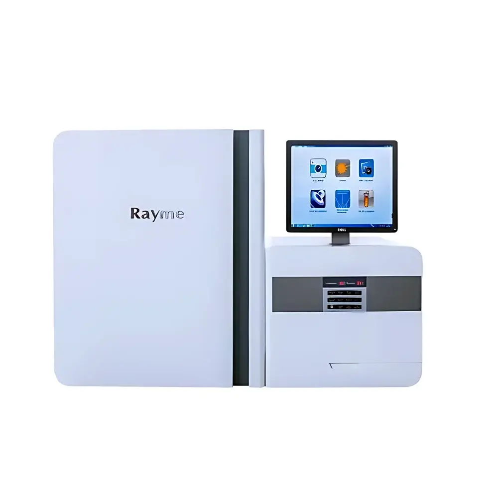
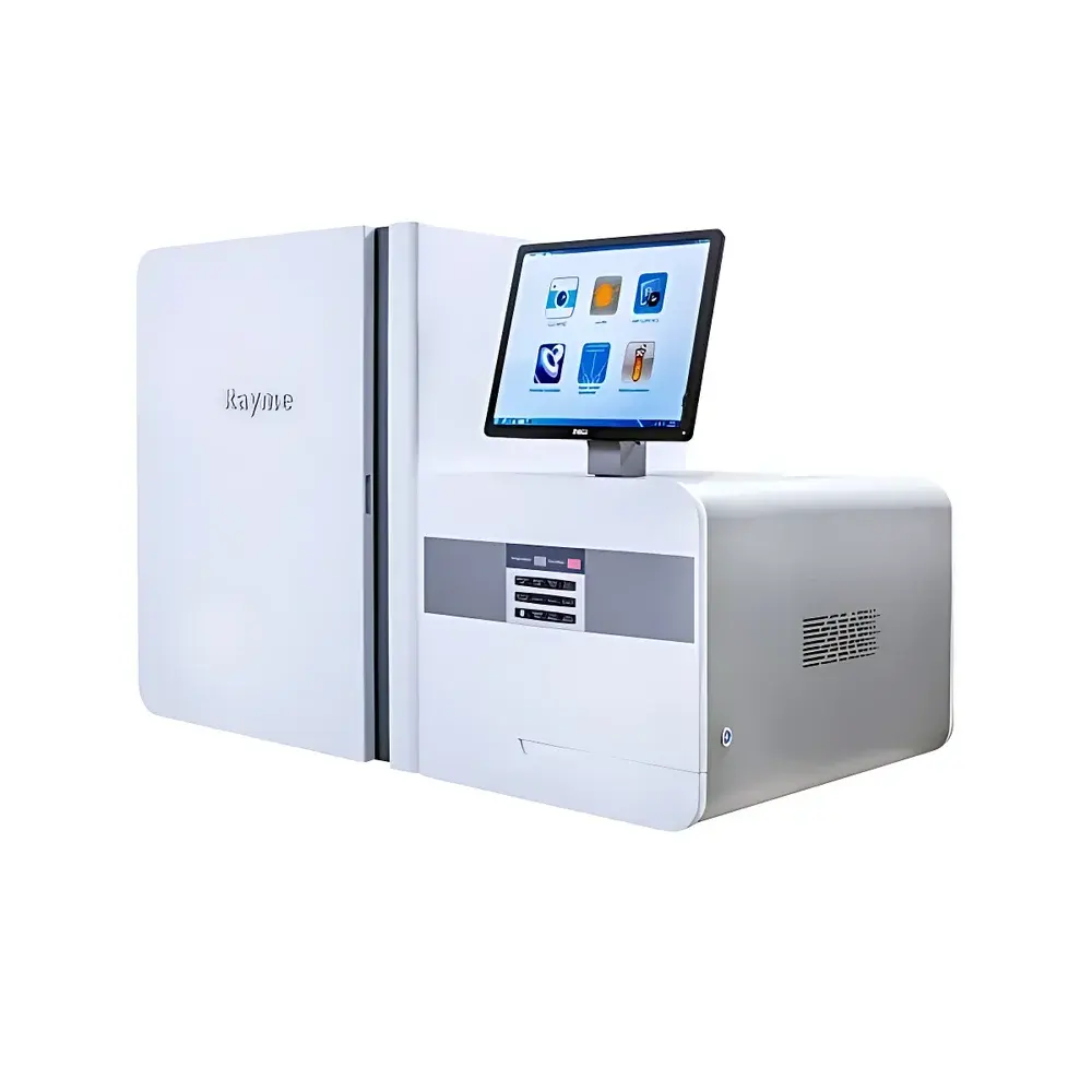
Raymetech SCA Series Real-Time In Situ Single-Cell Biochemical Analyzer
| Brand | Raymetech |
|---|---|
| Model | SCA Series |
| Origin | Jiangsu, China |
| Instrument Type | Real-time in situ single-cell biochemical analyzer |
| Sample Format | Live cells, tissue sections, in vivo preparations |
| Detection Modalities | Electrochemical, optical (fluorescence/absorbance), and hybrid dual-signal readout |
| Compliance Context | Designed for GLP-compliant research environments |
| Software Interface | Windows-based acquisition and analysis suite with timestamped metadata logging, ROI-based quantification, and kinetic trace export (CSV, HDF5) |
Overview
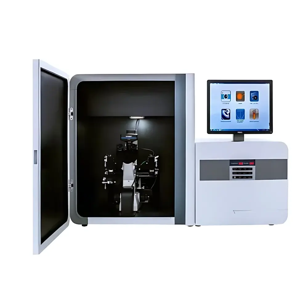
The Raymetech SCA Series Real-Time In Situ Single-Cell Biochemical Analyzer is an advanced platform engineered for label-free, non-destructive, and spatially resolved biochemical interrogation of individual living cells under physiological conditions. It integrates microelectrode-based amperometric/voltammetric sensing with high-sensitivity optical detection—typically fluorescence or absorbance—to enable concurrent measurement of enzymatic activity, redox state, neurotransmitter release, ion flux (e.g., Ca²⁺, H⁺, K⁺), metabolite secretion (e.g., glucose, lactate, dopamine, 5-HT), and nucleic acid hybridization events—all without requiring cell lysis or fixation. Unlike conventional bulk assays or endpoint fluorescence microscopy, the SCA system operates on the principle of localized electrochemical catalysis coupled with evanescent-field optical excitation, permitting subcellular spatial resolution (≤2 µm lateral) and millisecond temporal resolution. Its probe architecture supports both submerged culture and in vivo insertion, making it suitable for adherent monolayers, 3D organoids, ex vivo tissue slices, and awake-behaving rodent models via chronically implanted microsensors.
Key Features
- Real-time kinetic profiling: Continuous acquisition at up to 100 Hz sampling rate for dynamic biochemical events (e.g., enzyme turnover, oxidative burst, synaptic vesicle release)
- In situ compatibility: Operates directly within standard CO₂ incubators, laminar flow hoods, or stereotaxic rigs without compromising sterility or environmental control
- Multi-modal transduction: Simultaneous electrochemical + optical signal capture from identical cellular microdomains using co-localized microelectrodes and fiber-optic waveguides
- Minimal perturbation design: Ultra-microfabricated probes (tip diameter <5 µm) reduce mechanical stress and preserve native cell morphology and function
- Modular probe interface: Interchangeable sensor cartridges calibrated per analyte class (e.g., NADH oxidase, acetylcholinesterase, monoamine oxidase, superoxide dismutase)
- Temperature- and pH-stabilized fluidics: Integrated micro-perfusion system maintains physiological osmolarity and buffering capacity during extended recordings (>4 hr)
Sample Compatibility & Compliance
The SCA Series accommodates diverse biological specimens including primary mammalian neurons, immune cells, cancer spheroids, zebrafish embryos, and murine brain slices. All sensor surfaces undergo ISO 10993-5 cytocompatibility validation, and probe sterilization follows ANSI/AAMI ST79 guidelines. Data acquisition workflows comply with Good Laboratory Practice (GLP) documentation standards, including electronic signature support, user-access controls, and immutable audit trails. When deployed with Raymetech’s validated SCALink™ software (v4.2+), the system meets functional requirements for 21 CFR Part 11 compliance—including electronic record integrity, operator authentication, and change history logging—facilitating regulatory submissions in preclinical pharmacology and toxicology studies.
Software & Data Management
SCALink™ software provides synchronized multi-channel visualization, baseline correction algorithms (e.g., polynomial drift removal, adaptive noise filtering), and kinetic modeling tools (Michaelis–Menten fitting, Hill equation regression). Raw data are stored in HDF5 format with embedded metadata (sensor ID, calibration date, environmental parameters, user annotations). Export options include time-resolved CSV matrices, annotated TIFF stacks for co-registered imaging, and MATLAB-compatible .mat files. The software supports batch processing across ≥100 cells per experiment and integrates with third-party platforms such as ImageJ/Fiji (via Bio-Formats), MATLAB, and Python (via PySCA API). All data files retain cryptographic hash signatures for integrity verification.
Applications
- Pharmacology & Toxicology: Quantification of drug-induced mitochondrial uncoupling, cytochrome P450 metabolism kinetics, and off-target ion channel modulation at single-cell resolution
- Oncology Research: Mapping metabolic heterogeneity in tumor microenvironments, detecting early apoptosis via caspase-3 activity gradients, and evaluating PD-L1 expression dynamics in response to checkpoint inhibitors
- Neuroscience: Subsecond detection of dopamine efflux in striatal synaptosomes, real-time monitoring of glutamate spillover in hippocampal CA1, and longitudinal tracking of 5-HT release in dorsal raphe during behavioral assays
- Stem Cell Biology: Correlating glycolytic flux with pluripotency marker expression during induced differentiation, and resolving lineage-specific lactate dehydrogenase isoform switching
- Immunometabolism: Measuring real-time NADPH oxidase (NOX2) activity in phagocytosing macrophages and linking ROS production kinetics to cytokine secretion profiles
FAQ
What sample preparation protocols are required prior to analysis?
No fixation, permeabilization, or genetic labeling is needed. Cells are cultured on standard TC-treated substrates or embedded in low-melting-point agarose for suspension cultures. Tissue slices are maintained in oxygenated ACSF at 32°C.
Can the system distinguish between intracellular and extracellular analyte signals?
Yes—through differential probe positioning (intracellular impalement vs. pericellular placement) and selective membrane-impermeant enzyme substrates (e.g., AM-ester blocked fluorophores hydrolyzed only by cytosolic esterases).
Is calibration traceable to NIST standards?
Electrochemical sensors are factory-calibrated against certified reference materials (CRM) for dopamine, H₂O₂, and glucose (NIST SRM 2973, 2974); optical channels are validated using NIST-traceable fluorescence intensity standards (SRM 2940).
Does the system support long-term time-lapse experiments?
Yes—continuous operation up to 12 hours is supported with active thermal regulation, CO₂ compensation, and automated focus stabilization via piezoelectric objective control.
Are consumables proprietary or compatible with third-party electrodes/fibers?
Probe cartridges are Raymetech-proprietary, but the analog I/O interface complies with IEEE 1394b and supports integration with custom-built microelectrodes or commercial optical fibers (e.g., Thorlabs, Oz Optics) via adapter modules.


