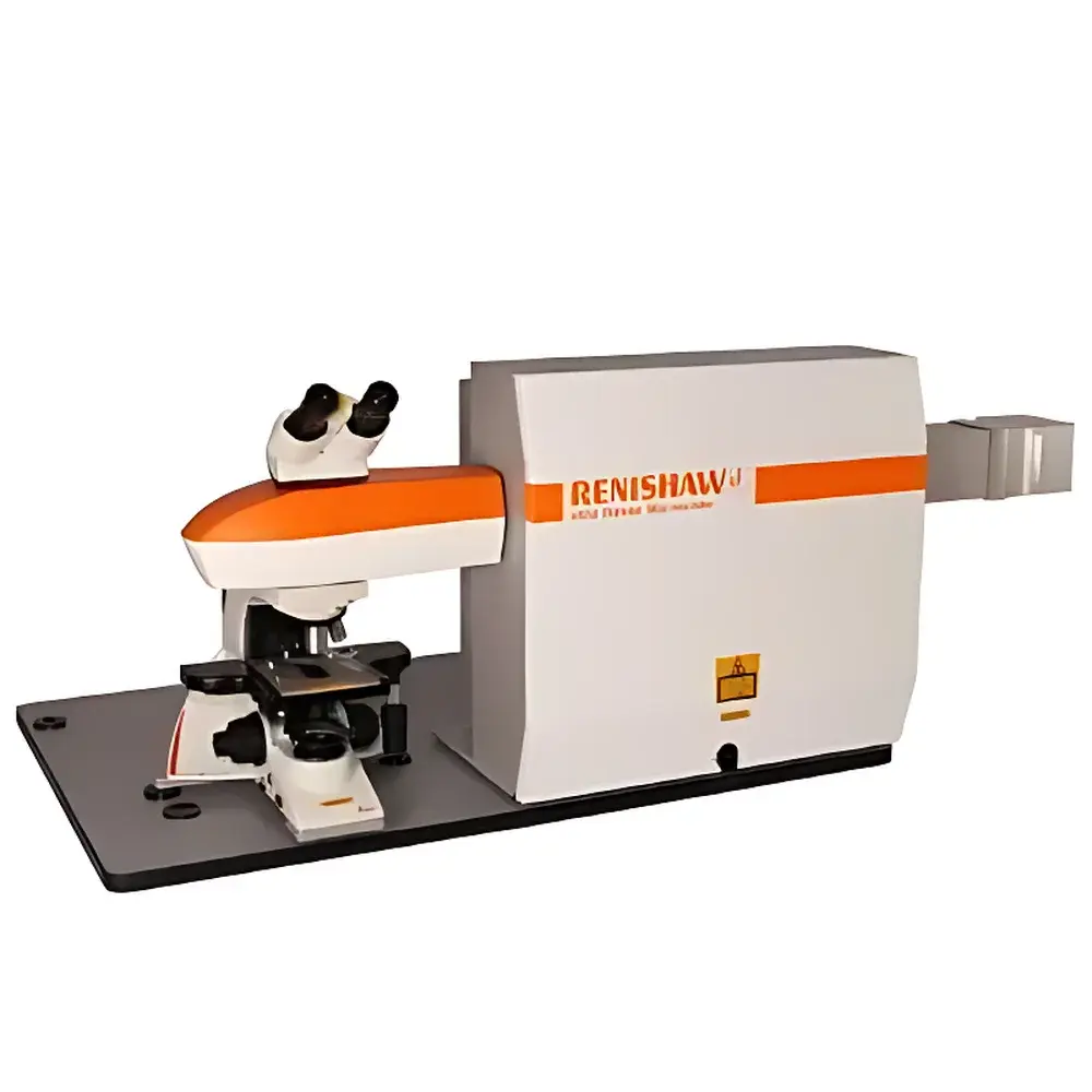
Renishaw inVia Reflex Confocal Raman Microscope
| Brand | Renishaw |
|---|---|
| Origin | United Kingdom |
| Model | inVia Reflex |
| Instrument Type | Confocal Raman Microscope |
| Spectral Range | 100–4000 cm⁻¹ |
| Spectral Resolution | ≤1 cm⁻¹ |
| Spatial Resolution | XY ≈ 1 µm, Z ≈ 2 µm |
| Low-Wavenumber Limit | 10 cm⁻¹ |
| Spectral Reproducibility | ≤0.1 cm⁻¹ |
Overview
The Renishaw inVia Reflex Confocal Raman Microscope is a research-grade, fully automated confocal Raman spectroscopy platform engineered for high-precision molecular characterization at the micro- and sub-micron scale. Based on confocal optical architecture and laser-induced Raman scattering, the system delivers spatially resolved vibrational spectra by selectively collecting signal from a defined focal volume—rejecting out-of-focus emission through a pinhole aperture. This principle enables depth-sectioning, 3D chemical mapping, and quantitative analysis of heterogeneous samples with minimal interference from substrate or overlying layers. Designed and manufactured in the UK, the inVia Reflex integrates Renishaw’s decades-long expertise in precision optomechanics, metrology-grade motion control, and spectral calibration traceability. Its modular architecture supports multi-laser excitation (UV–VIS–NIR), interchangeable gratings, and real-time spectral optimization—making it suitable for academic research, industrial R&D, and regulated quality control environments where reproducibility, long-term stability, and audit-ready documentation are essential.
Key Features
- Fully automated operation: motorized selection of lasers, edge filters, and diffraction gratings; self-aligning optical path; automated wavelength calibration and intensity normalization
- Confocal design with diffraction-limited spatial resolution: XY ≈ 1 µm (dependent on objective NA and excitation wavelength), axial (Z) resolution ≈ 2 µm
- High spectral fidelity: ≤1 cm⁻¹ resolution across 100–4000 cm⁻¹ range; low-wavenumber capability down to 10 cm⁻¹ using optimized notch filters and high-efficiency optics
- Multi-detector configuration: supports up to four back-illuminated, deep-depletion CCD detectors for simultaneous multi-spectral acquisition or extended range coverage
- Research-grade microscope integration: compatible with both upright and inverted configurations; supports Köhler illumination, DIC, fluorescence, and polarization contrast
- Robust mechanical architecture: granite optical base, air-bearing translation stages (optional), and thermal drift compensation ensure measurement stability over extended acquisition times
Sample Compatibility & Compliance
The inVia Reflex accommodates diverse sample types—including thin films, powders, biological tissues, polymers, semiconductors, pharmaceuticals, and geological specimens—without destructive preparation. Optional environmental modules (e.g., cryostats, heating stages, electrochemical cells, and humidity-controlled chambers) enable in situ and operando measurements under non-ambient conditions. The system complies with international standards relevant to analytical instrumentation: ISO/IEC 17025 requirements for calibration traceability, ASTM E1840 and E2598 for Raman spectral validation, and USP for pharmaceutical solid-state characterization. When configured with audit-trail-enabled software and electronic signatures, it supports GLP and GMP workflows compliant with FDA 21 CFR Part 11.
Software & Data Management
Controlled by Renishaw’s WiRE™ (Witec Raman Environment) software, the inVia Reflex provides an integrated environment for instrument control, spectral acquisition, multivariate analysis (PCA, cluster analysis, spectral unmixing), and hyperspectral imaging. WiRE supports scripting (Python API), batch processing, and metadata-rich data storage compliant with AnIML and JCAMP-DX formats. All calibrations, alignment logs, and maintenance records are automatically timestamped and archived. Raw spectral data, processed maps, and report templates are exportable in open formats (CSV, HDF5, TIFF) for third-party analysis or LIMS integration. Software updates are delivered via secure channels with version-controlled release notes and backward-compatible file handling.
Applications
- Materials science: phase identification in battery cathodes, stress/strain mapping in 2D materials (e.g., graphene, MoS₂), crystallinity assessment in polymers and thin-film solar cells
- Life sciences: label-free cellular imaging, lipid/protein distribution in tissue sections, drug–excipient interactions in solid dispersions
- Pharmaceuticals: polymorph screening, counterfeit detection, content uniformity mapping in tablets, stability-indicating assays
- Geosciences: mineral assemblage analysis, fluid inclusion characterization, carbon speciation in metamorphic rocks
- Nanotechnology: plasmonic nanoparticle identification, surface-enhanced Raman spectroscopy (SERS) substrate validation, nanocomposite homogeneity assessment
- Forensics: pigment and dye identification in inks, paints, and fibers; explosive residue detection; microplastic classification
FAQ
What laser wavelengths are supported on the inVia Reflex?
Standard configurations include 325 nm, 532 nm, 633 nm, and 785 nm lasers; additional wavelengths (e.g., 405 nm, 830 nm) are available as options.
Can the system perform correlative analysis with other techniques?
Yes—the inVia Reflex features standardized mechanical and electrical interfaces for seamless integration with AFM, SEM, CLSM, and photoluminescence systems via Renishaw’s Correlate™ platform.
Is spectral calibration traceable to NIST standards?
Yes—each system ships with factory-calibrated reference spectra (e.g., silicon, cyclohexane, sulfur) and optional NIST-traceable calibration kits for on-site verification.
How is data integrity ensured during long-duration mapping experiments?
WiRE software implements real-time checksum validation, automatic backup intervals, and hardware-triggered acquisition synchronization to prevent data loss or misalignment.
Does the system support automated particle analysis?
Yes—WiRE’s Particle Analysis module enables size, shape, and chemical classification of individual particles within Raman image stacks, with configurable thresholds and statistical reporting.
Related Products


")