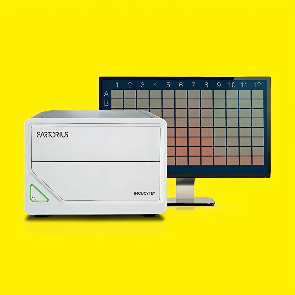
Sartorius Incucyte® S3/SX1/SX5 Live-Cell Analysis System
| Brand | Sartorius |
|---|---|
| Origin | USA |
| Model | Incucyte S3, Incucyte SX1, Incucyte SX5 |
| Imaging Modes | Phase Contrast, Green Fluorescence (e.g., GFP), Red Fluorescence (e.g., RFP/mCherry) |
| Objective Lenses | 4×, 10×, 20× motorized auto-focus |
| Throughput | Up to 6 multiwell plates (96-well or 384-well) per run |
| Environmental Integration | Designed for continuous operation inside standard CO₂ incubators (37 °C, 5% CO₂, humidified) |
| Software Platform | Incucyte® Base Software v2023.1+ with VesselView™, PlateGraph™, and Mask-based analysis modules |
| Compliance | Supports audit trails, user access control, and electronic signatures compliant with FDA 21 CFR Part 11 and GLP/GMP workflows |
| Data Output | Time-lapse image stacks, quantitative metrics (confluence %, object count, neurite length, spheroid area/volume, migration distance), exportable to CSV, PNG, TIFF, and PDF |
Overview
The Sartorius Incucyte® S3, SX1, and SX5 Live-Cell Analysis Systems are integrated, incubator-adapted imaging platforms engineered for label-free and fluorescent kinetic monitoring of mammalian cells under physiologically relevant conditions. Leveraging widefield microscopy with motorized optics and environmental stability—no sample removal from the incubator is required—the system captures high-resolution phase contrast and dual-channel fluorescence (typically 470/525 nm and 560/620 nm) time-lapse data over hours to weeks. Its core measurement principle relies on automated, repeated optical sectioning across defined positions within culture vessels, followed by algorithm-driven segmentation and quantification of morphological, proliferative, migratory, and functional cellular parameters. Unlike endpoint assays or plate readers, the Incucyte platform delivers longitudinal, context-preserving datasets that reflect true biological dynamics—critical for mechanistic studies in oncology, immunology, neuroscience, and regenerative medicine.
Key Features
- Motion-stabilized optical design: The objective and illumination module move while samples remain stationary—minimizing mechanical disturbance to sensitive or non-adherent cells (e.g., suspension lymphocytes, organoids, or spheroids).
- Multi-objective flexibility: Switch between 4× (broad-field confluence), 10× (subcellular morphology), and 20× (fine structural detail) objectives without manual intervention; all calibrated for consistent magnification and depth-of-field across experiments.
- Incubator-native architecture: Fully sealed, vibration-damped chassis rated for continuous operation at 37 °C and 5% CO₂; compatible with standard tissue culture incubators via pass-through cable routing or wireless Ethernet.
- Modular assay compatibility: Pre-validated reagent kits—including NucLight™ nuclear labeling, Caspase-3/7 apoptosis sensors, pHrodo™ phagocytosis probes, and ClearView™ chemotaxis plates—are optimized for minimal cytotoxicity and maximal signal-to-noise ratio.
- Multi-user, multi-protocol workflow support: Role-based login, independent acquisition schedules per user, and concurrent analysis of heterogeneous plate formats (e.g., 96-well for dose-response, 384-well for high-throughput screening) within a single instrument session.
Sample Compatibility & Compliance
The Incucyte systems accept standard cell culture formats including T-flasks, Petri dishes, and multiwell plates (6–384-well), with full support for both adherent and suspension models. Applications span primary human cells, iPSC-derived lineages, co-cultures (e.g., tumor–T cell, neuron–astrocyte), 3D spheroids, and microfluidic chips. All hardware and software components comply with ISO 13485:2016 (medical device quality management) and meet technical requirements for regulated environments: audit trail logging (user actions, parameter changes, analysis versioning), electronic signature enforcement, and secure data encryption (AES-256). The Base Software is validated for use in GLP-compliant toxicology studies and GMP-aligned bioprocess development workflows per ICH M10 and USP .
Software & Data Management
Incucyte Base Software provides an intuitive, wizard-guided interface for experiment setup, image acquisition scheduling, and batch analysis. Core modules include VesselView™ (real-time visualization of entire wells with region-of-interest masking), PlateGraph™ (multi-parameter trend plotting across all wells), and custom algorithm scripting via Python API integration. Raw image data (16-bit TIFF stacks) and derived metrics are stored in a local SQL Server database with optional cloud backup. Export formats include publication-ready figures (SVG/PNG), statistical summaries (CSV/Excel), and FAIR-compliant metadata (MIAME/MINSEQE-aligned). Version-controlled analysis pipelines ensure reproducibility across labs and longitudinal studies.
Applications
- Cell Health & Viability: Label-free confluence tracking, NucLight™-based proliferation kinetics, Caspase-3/7 activation for apoptosis, and ATP-independent cytotoxicity profiling.
- 3D Models: Automated spheroid segmentation, growth curve modeling, necrotic core detection, and drug-induced disintegration quantification.
- Immunooncology: Real-time tumor cell killing by CAR-T/NK cells, immune synapse formation, and phagocytic index calculation using pHrodo™-labeled targets.
- Neuroscience: Neurite outgrowth dynamics, branching complexity scoring, and synaptic puncta colocalization in co-culture systems.
- Migration & Invasion: Scratch wound closure kinetics, transwell-free chemotaxis assessment in ClearView™ plates, and matrix metalloproteinase-dependent invasion through collagen gels.
- Functional Genomics: Reporter gene expression (GFP/RFP), CRISPR-edited clone validation, and transfection efficiency mapping across 96-well arrays.
FAQ
Can the Incucyte system be used outside a CO₂ incubator?
Yes—while optimized for incubator-integrated operation, the S3/SX1/SX5 models support ambient-air imaging with external humidity control units for short-term assays requiring normoxic or hypoxic conditions.
What level of image resolution is achieved at 20× magnification?
At 20×, the system delivers effective pixel resolution of ≤1.2 µm with diffraction-limited contrast; lateral resolution is validated per ISO 19012-1 using USAF 1951 test targets.
Is remote access supported for instrument control and data review?
All models include browser-based remote dashboard access via HTTPS; licensed users can initiate acquisitions, adjust focus, and launch analyses from any network-connected device without VPN configuration.
How does the system handle focus drift during long-term imaging?
Each acquisition cycle includes Z-stack autofocus using contrast maximization algorithms; reference focus maps are updated hourly or triggered by environmental fluctuation detection.
Are analysis algorithms validated against gold-standard methods?
Yes—Sartorius provides analytical method validation reports (AMVRs) demonstrating correlation with flow cytometry (proliferation/apoptosis), manual counting (spheroid area), and transwell assays (migration), with r² ≥0.98 across >50 independent benchmarks.


