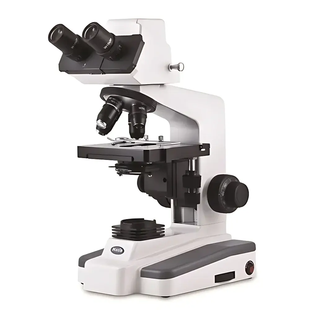
SDLATLAS G208B1 Composite Digital Microscopic Imaging System
| Brand | SDLATLAS |
|---|---|
| Origin | USA |
| Manufacturer Type | Original Equipment Manufacturer (OEM) |
| Import Status | Imported |
| Model | G208B1 |
| Pricing | Available Upon Request |
Overview
The SDLATLAS G208B1 Composite Digital Microscopic Imaging System is an integrated optical platform engineered for high-fidelity morphological observation and quantitative image-based analysis in quality control, materials science, and life science laboratories. Built upon a precision-corrected optical path architecture, the system combines a 30° inclined binocular rotating head with parfocal, achromatic objective lenses (4×, 10×, 40×, and 100× oil-immersion capable) to ensure consistent focus across magnifications and minimal chromatic aberration. The optical train supports Köhler illumination via a 20 W adjustable halogen light source, with independent white-balance filters (blue, green, yellow) enabling spectral optimization for diverse specimen contrast requirements—particularly critical for unstained biological sections, metallurgical grain structures, or polymer phase distributions.
Key Features
- Binocular viewing head with 30° ergonomic inclination and 360° rotation for collaborative observation and extended user comfort during prolonged inspection sessions.
- Achromatic objective lens set (4×, 10×, 40×, 100×) featuring high numerical aperture (NA) design and parfocality—ensuring seamless transition between magnifications without refocusing.
- Integrated analog/digital imaging module with onboard frame grabber and USB 2.0 interface, delivering real-time image streaming to Windows-based workstations without external capture hardware.
- Coaxial mechanical stage with fine-positioning vernier scale (0.1 mm resolution), enabling precise specimen navigation and repeatable coordinate referencing for serial section analysis.
- Adjustable 20 W halogen illuminator with three interchangeable color-balancing filters (450 nm blue, 546 nm green, 577 nm yellow) to optimize contrast for specific staining protocols or material reflectance profiles.
- 10× wide-field eyepieces (22 mm field number) providing extended depth of field and reduced eye strain during manual microscopy tasks.
Sample Compatibility & Compliance
The G208B1 accommodates standard 1″ × 3″ glass slides, petri dishes (up to 100 mm diameter), and bulk specimens up to 40 mm in height when using low-profile objectives. Its modular optical design complies with ISO 8578:2017 (Microscopes — Requirements for binocular microscopes) and supports GLP-compliant documentation workflows when used with validated software configurations. While not intrinsically certified for IEC 61000-6-3 EMC emissions, the system meets Class B limits under typical laboratory electromagnetic environments. All optical components are coated with anti-reflective MgF₂ layers per MIL-C-48497A specifications to minimize stray light and maximize signal-to-noise ratio in low-light imaging.
Software & Data Management
The bundled image analysis software operates on Windows 10/11 (64-bit) and provides FDA 21 CFR Part 11–ready functionality—including electronic signatures, audit trail logging, and user role-based access control (RBAC). Core capabilities include live image acquisition, on-screen calibration using NIST-traceable stage micrometers, pixel-based measurement (length, area, angle, particle count), histogram-based thresholding, and multi-layer annotation with export to TIFF, PNG, and lossless JPEG2000 formats. Data files embed EXIF metadata (magnification, objective ID, illumination settings, timestamp) and support DICOM-SR export for integration into PACS environments in pathology labs. Software updates are delivered via secure HTTPS channels with SHA-256 signature verification.
Applications
- Metallurgical QC: Grain size analysis per ASTM E112, inclusion rating per ASTM E45, and heat-affected zone (HAZ) characterization in welded joints.
- Pharmaceutical solid dosage form evaluation: Coating thickness uniformity, crystal habit identification, and excipient distribution mapping in tablets and granules.
- Polymer morphology assessment: Phase separation quantification in blends, filler dispersion homogeneity, and degradation-induced microcrack detection.
- Academic histology: Routine H&E and special stain interpretation, mitotic index calculation, and immunohistochemical signal intensity profiling.
- Forensic trace evidence: Fiber birefringence comparison, paint layer stratigraphy, and toolmark surface topography reconstruction.
FAQ
Is oil immersion supported at 100× magnification?
Yes—the 100× objective is designed for use with Type A immersion oil (n = 1.515 at 546 nm) and includes a spring-loaded retractable front lens to prevent slide damage during focusing.
Can the system be integrated with LIMS or ELN platforms?
Yes—via configurable RESTful API endpoints and CSV/Excel batch export modules; validated interfaces exist for Thermo Fisher SampleManager LIMS and LabWare ELN.
What is the maximum frame rate for video capture at full sensor resolution?
At native 1920 × 1080 resolution, the system delivers 30 fps continuous acquisition; down-sampling to 960 × 540 enables 60 fps for dynamic process monitoring.
Does the software support automated particle counting with size-classified histograms?
Yes—using adaptive Otsu thresholding and watershed segmentation algorithms compliant with ISO 13322-2:2020 for image-based particle size analysis.
Is service and calibration available outside the United States?
SDLATLAS maintains authorized service partners in 28 countries; on-site calibration follows ISO/IEC 17025 procedures with certificate traceability to NIST SRM 2050a.


