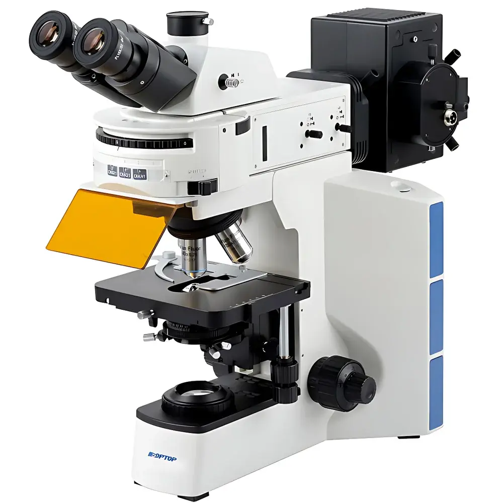
SOPTOP CX40 Upright Fluorescence Microscope
| Brand | SOPTOP |
|---|---|
| Origin | Zhejiang, China |
| Manufacturer Type | Original Equipment Manufacturer (OEM) |
| Product Category | Domestic |
| Model | CX40 |
| Instrument Type | Upright Fluorescence Microscope |
| Excitation Source | High-Efficiency Multi-Wavelength LED System (UV/B/G) |
| Illumination | 6V30W Halogen Lamp (Centered, Continuously Adjustable Intensity) |
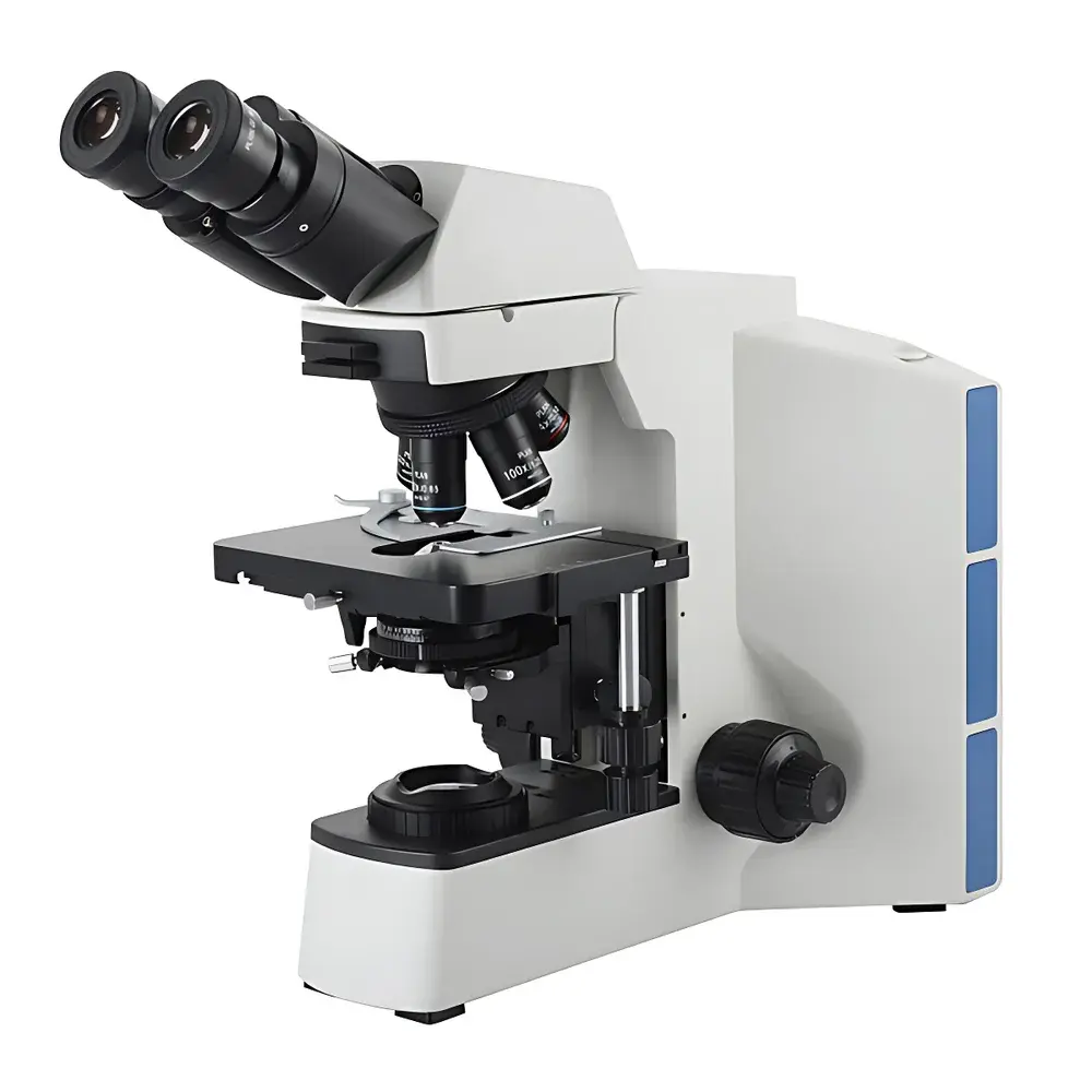
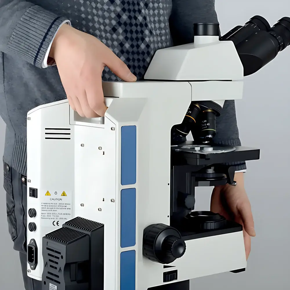
| Observation Head | Infinity-Corrected Trinocular Hinged Tube (Optional Binocular) |
| Eyepieces | Wide-Field Plan Eyepieces PL10X/22mm with Optional Reticle |
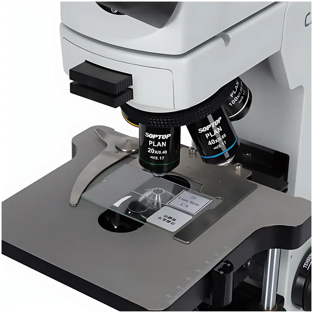
| Objectives | Infinity-Corrected Achromatic (2X–100X), Phase Contrast (10X–100X), and Semi-Apochromatic Fluor (4X–100X) Objective Sets |
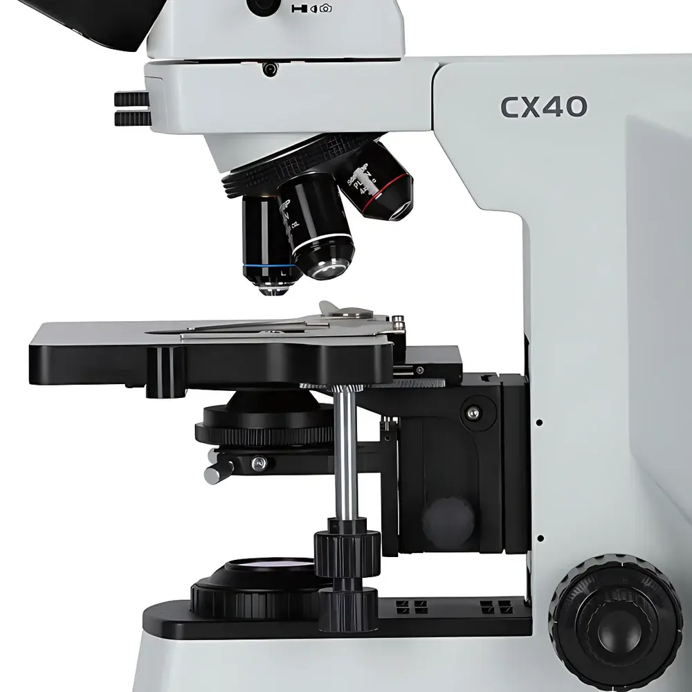
| Condenser | Swing-Out Achromatic Condenser, NA 1.2/0.22, Graduated Iris Diaphragm |
| Stage | 175 × 145 mm Right-Hand Dual-Layer Mechanical Stage (76 × 50 mm Travel, 0.1 mm Resolution) |
| Fluorescence Filter Sets | UV1/V1/B1/G1 |
| DIC Compatibility | Built-in DIC Slots in Nosepiece & Condenser |
| Focusing Mechanism | Low-Position Coaxial Coarse/Fine Focus with 1 µm Fine Adjustment Graduation |
| Control Mode | Manual |
| Regulatory Status | Not a Medical Device (Non-CE/IEC 62304, Non-FDA 510(k)) |
Overview
The SOPTOP CX40 Upright Fluorescence Microscope is an engineered platform for high-fidelity epifluorescence imaging in life science laboratories, clinical pathology support environments, and academic research settings. Designed around an infinity-corrected optical architecture, the system integrates multi-band LED excitation (UV, violet, blue, and green), semi-apochromatic PLAN-FLUOR fluor objectives, and precision mechanical staging to deliver consistent signal-to-noise ratios across fluorescence modalities—including DAPI, FITC, TRITC, and Cy3/Cy5 analogues. Its Y-shaped monolithic aluminum alloy frame provides structural rigidity exceeding 0.5 µm lateral stability at 100× oil immersion—critical for multi-channel registration in FISH, immunofluorescence co-localization, and time-lapse z-stack acquisition. Unlike mercury arc lamp systems, the CX40’s solid-state LED illumination eliminates warm-up delays, thermal drift, and ozone generation while offering >100,000 hours of operational lifetime and continuous intensity modulation without spectral shift.
Key Features
- Modular LED epifluorescence illumination with synchronized filter turret—enabling seamless switching between UV1, V1, B1, and G1 filter cubes without realignment
- Infinity-corrected semi-apochromatic PLAN-FLUOR objectives (4X–100X) featuring enhanced transmission in UV range (250–400 nm) and optimized spherical/chromatic aberration correction across visible spectrum
- Dual-layer mechanical stage with 76 × 50 mm travel, 0.1 mm vernier resolution, and reversible right/left-hand configuration; optional ceramic-coated variant resists corrosion and minimizes thermal expansion-induced drift
- Swing-out achromatic condenser (NA 1.2/0.22) with calibrated iris diaphragm and built-in phase contrast annuli for simultaneous brightfield/phase/fluorescence workflows
- Trinocular observation head with 20° inclined eyepieces (PL10X/22 mm), optional reticle integration, and C-mount port for standardized camera coupling (e.g., USB3.0 sCMOS or EMCCD)
- DIC-ready design: nosepiece includes dedicated slots for Wollaston prisms; condenser supports centerable Nomarski prism insertion for quantitative optical path difference imaging
Sample Compatibility & Compliance
The CX40 accommodates standard glass slides (1 × 3 inches), coverslips (No. 1.5, 0.17 mm thickness), and thick-sectioned tissue specimens up to 100 µm in depth under oil immersion. Its mechanical stage permits dual-slide comparison—enabling side-by-side analysis of control vs. experimental sections during diagnostic screening. While not certified as a medical device per ISO 13485 or FDA 21 CFR Part 820, the microscope conforms to IEC 61000-6-3 (EMC emissions) and IEC 61000-6-2 (immunity). All optical components comply with ISO 8578 (microscope objective labeling) and ISO 10934-1 (image quality testing protocols). The LED excitation system meets IEC 62471 photobiological safety Class 1 requirements for both UV and visible bands.
Software & Data Management
The CX40 operates as a hardware platform compatible with third-party imaging software suites—including NIS-Elements (Nikon), ZEN Blue (Zeiss), and open-source platforms such as MicroManager and Fiji/ImageJ—via standard USB 3.0 or GigE Vision interfaces. No proprietary acquisition software is bundled; instead, the system delivers raw 12-bit or 16-bit TIFF streams with embedded metadata (objective ID, magnification, filter position, exposure time). Audit trails, user access logs, and electronic signatures are managed externally through laboratory information management systems (LIMS) compliant with GLP and 21 CFR Part 11 when paired with validated middleware.
Applications
- Fluorescence in situ hybridization (FISH) assay validation and enumeration in hematopathology and cytogenetics labs
- Immunofluorescent detection of intracellular protein localization and post-translational modifications
- Live-cell imaging support (with environmental chamber integration) using low-phototoxicity LED excitation
- Multi-modal histopathology review combining brightfield H&E, phase contrast for unstained specimens, and fluorescence for biomarker verification
- Teaching laboratories requiring robust, serviceable instrumentation for undergraduate microscopy instruction and graduate-level advanced imaging techniques
FAQ
Is the CX40 suitable for quantitative fluorescence intensity measurements?
Yes—when used with calibrated photometric cameras and standardized exposure protocols, the stable LED output and high-transmission filter sets enable reproducible relative intensity comparisons across samples.
Can the system be upgraded to support motorized Z-axis or automated filter switching?
No—the CX40 is a manually operated platform with fixed mechanical controls; motorization is not supported by design.
What is the maximum usable magnification with the 100X oil immersion objective?
With optimal Köhler illumination and NA-matched condenser settings, the practical resolution limit is ~200 nm laterally (per Abbe criterion), supporting routine subcellular feature discrimination.
Does SOPTOP provide application-specific training or installation support?
SOPTOP offers on-site basic setup and optical alignment verification; advanced technique training (e.g., DIC optimization or multi-channel registration) is available via authorized regional partners.
Are replacement LED modules field-serviceable?
Yes—LED light engines are modular and replaceable without optical recalibration; spare units ship with factory spectral calibration reports.


