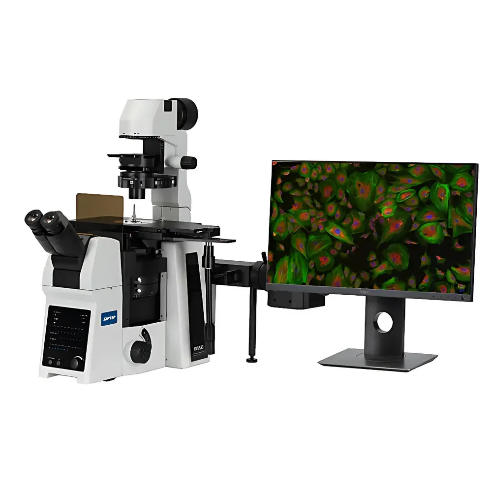
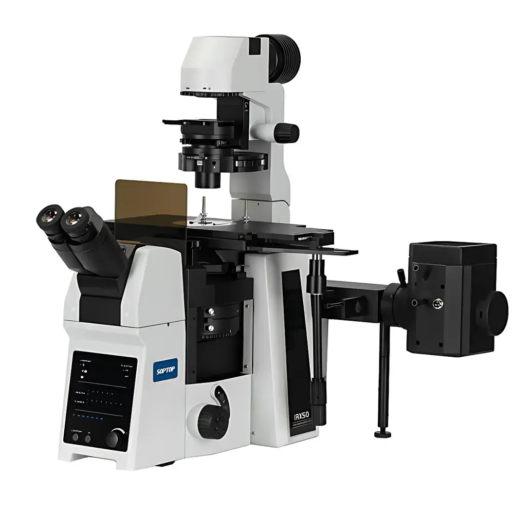
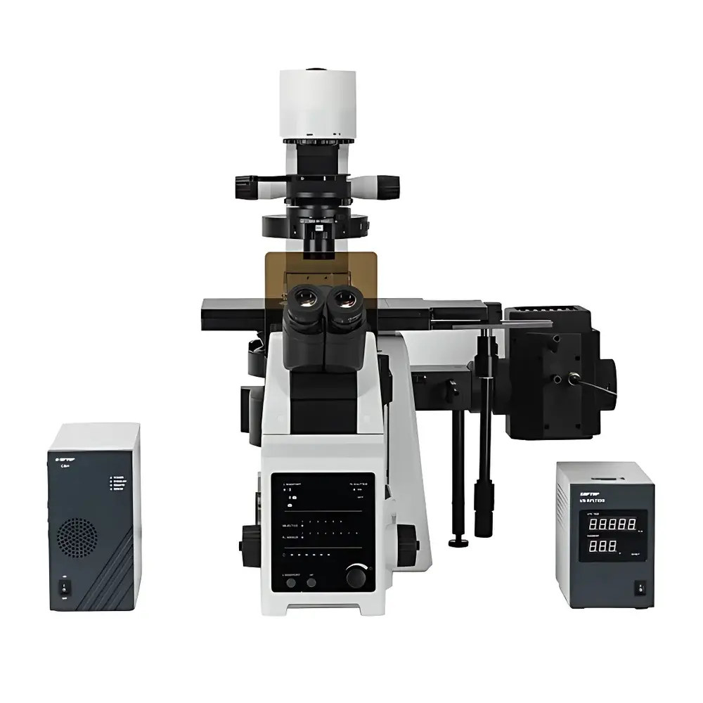
SOPTOP IRX50 Research-Grade Inverted Fluorescence Microscope
| Brand | SOPTOP |
|---|---|
| Origin | Zhejiang, China |
| Manufacturer Type | OEM Manufacturer |
| Product Category | Domestic |
| Model | IRX50 |
| Instrument Type | Inverted Fluorescence Microscope |
| Excitation Source | 100 W DC Mercury Arc Lamp (OSRAM) |
| Medical Device Classification | Not Applicable |
| Instrument Grade | Research-Grade |
| Eyepieces | High-Eyepoint Wide-Field Plan Eyepieces PL10X/22 mm with Adjustable Diopter |
| Objectives | Long Working Distance Semi-Apochromatic Plan Objectives (20X, 40X, 60X with Correction Collar) |
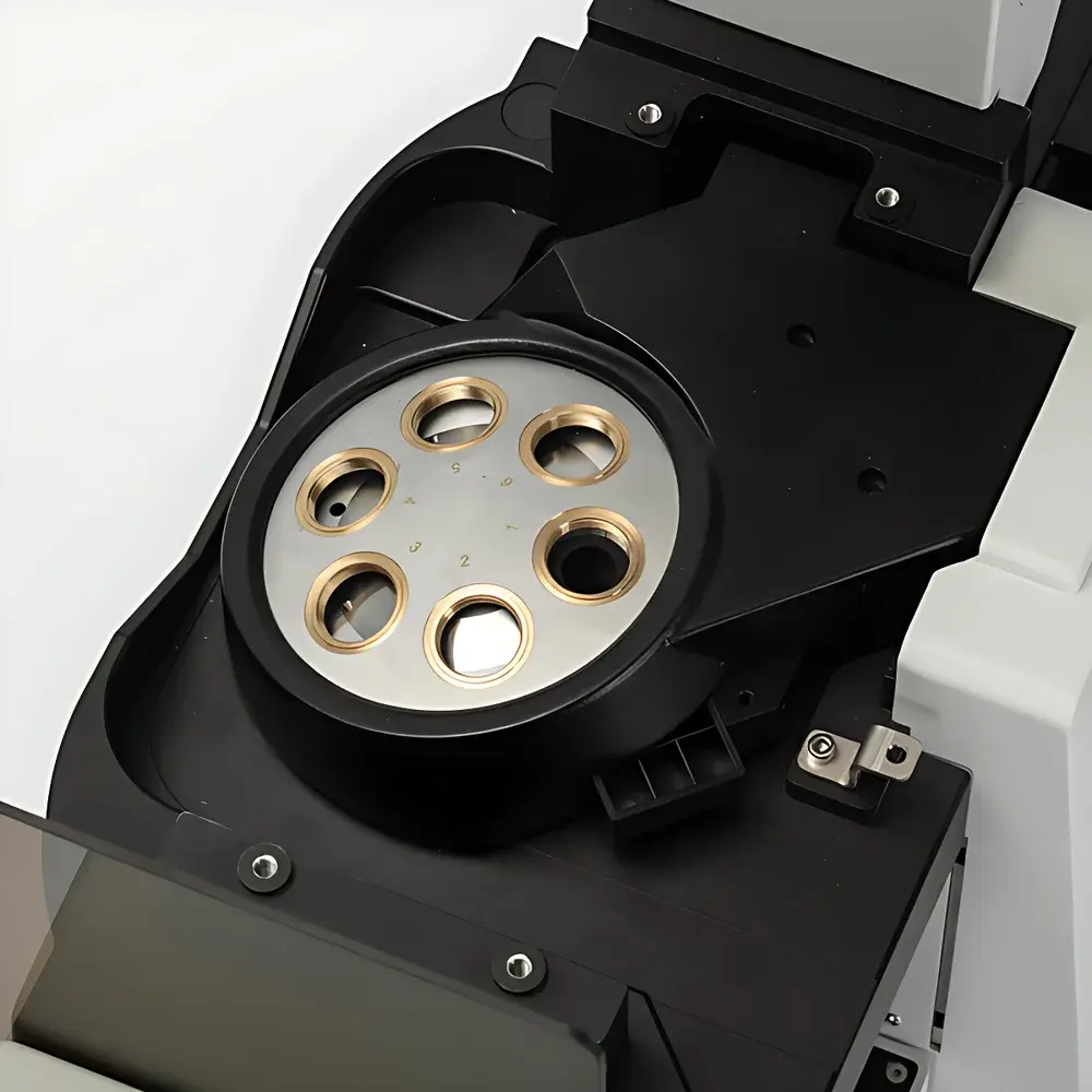
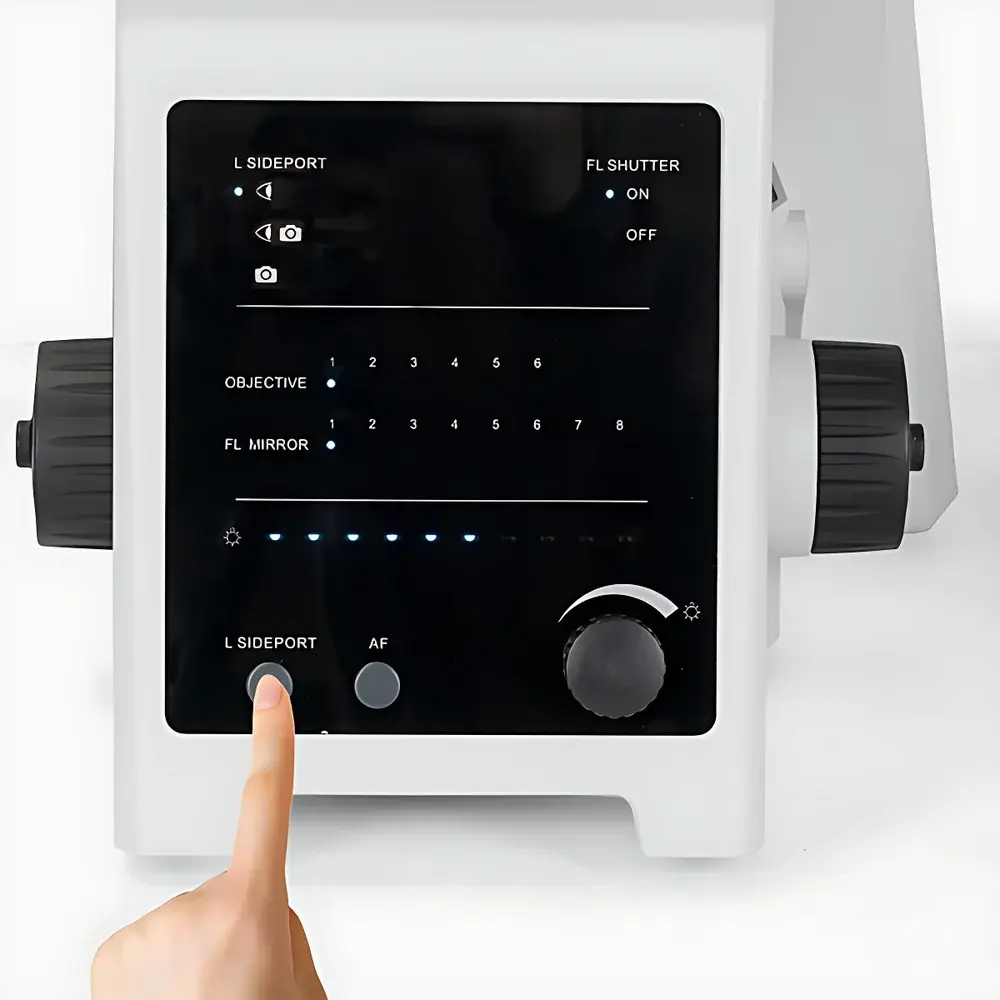
| Fluorescence Module | Encoded 8-Position Filter Turret (expandable to dual-layer, 16 total positions), motorized shutter with positional recognition (upper/lower layer detection) |
| Standard Filter Sets | B1 (EX480/30, DI505DC, EM535/40), G1 (EX560/40, DI600DC, EM635/60), UV1 (EX375/28, DI415DC, EM46/50), R1 |
| Illumination Systems | 100 W Mercury Lamp Housing + Power Supply + L-Type Fluorescence Illuminator with Attenuation Filter Slot |
| Control Mode | Motorized & Manual Hybrid |
| Focus Mechanism | Motorized Z-Axis Drive |
| Condenser | Manual 7-Position Condenser (NA 0.55, WD 27 mm) |
| Observation Head | Inclined Binocular Tube (20°–45° adjustable tilt), inverted image path, infinity-corrected, interpupillary distance 50–76 mm |
| DIC Components | Compatible via condenser and objective-side prism sets |
| Stage | Mechanical XY Stage (≥300 mm × 240 mm surface area), travel range 135 mm (X) × 85 mm (Y), thickness 30 mm |
| Fluorescence Filter Options | B/G/UV/R1 |
Overview
The SOPTOP IRX50 is a research-grade inverted fluorescence microscope engineered for high-fidelity live-cell imaging, developmental biology, and long-term time-lapse studies in academic and industrial life science laboratories. Built on an infinity-corrected optical architecture, the IRX50 leverages advanced Köhler illumination principles and precision-matched optical pathways to deliver consistent axial resolution, minimal chromatic aberration, and high signal-to-noise ratio across widefield fluorescence modalities. Its inverted configuration enables direct access to the specimen plane—ideal for observing adherent cells in standard Petri dishes, multi-well plates, and specialized culture chambers without mechanical obstruction. The system’s modular light-path design supports both single- and dual-layer beam-splitting configurations, facilitating integration with third-party accessories such as external cameras, spectrographs, or custom excitation sources while preserving optical throughput and alignment stability.
Key Features
- Motorized Precision Mechanics: Fully encoded Z-axis drive with sub-micron repeatability, motorized nosepiece with position memory, programmable filter turret with layer-aware positioning logic, and motorized port selector enable automated acquisition protocols compliant with GLP and ISO 17025 workflows.
- Optical Flexibility: Interchangeable condenser modules support brightfield, phase contrast, differential interference contrast (DIC), and relief contrast—each optimized for specific specimen thicknesses and refractive index gradients. The NA 0.55 condenser accommodates high-resolution DIC; the NA 0.3 ultra-long WD condenser ensures compatibility with thick-bottomed culture vessels and microfluidic devices.
- Fluorescence Optimization: The 8-position encoded filter turret accepts up to 16 filter sets across two stacked layers. Integrated motorized shutter provides precise exposure control, reducing phototoxicity during extended acquisitions. Standard B1, G1, UV1, and R1 filter sets cover common fluorophores including DAPI, FITC, TRITC, Cy5, and Hoechst.
- Ergonomic Design: Tiltable observation head (20°–45°), adjustable interpupillary distance (50–76 mm), and front-panel LCD status display reduce operator fatigue during prolonged imaging sessions. The low-profile joystick and XY lock mechanism enhance positional reproducibility for serial imaging of multiple fields-of-view.
- Objective Versatility: A full suite of long working distance semi-apochromatic and apochromatic objectives—including correction collar-equipped variants—compensates for spherical aberration induced by variable coverslip thicknesses (0.13–0.22 mm) and plastic-bottomed vessels, ensuring diffraction-limited performance across diverse sample formats.
Sample Compatibility & Compliance
The IRX50 accommodates standard and non-standard biological specimens including monolayer cultures, spheroids, organoids, zebrafish embryos, C. elegans, and tissue explants mounted in glass-bottom dishes (e.g., MatTek, Ibidi), chambered coverslips, and custom microfabricated substrates. Its ≥73 mm working distance objective options and 135 mm × 85 mm stage travel enable stable imaging of large-area samples and multi-position time-series experiments. The system complies with IEC 61000-6-3 (EMC emission standards) and IEC 61000-6-2 (immunity). While not classified as a medical device under FDA 21 CFR Part 809 or EU MDR Annex XVI, its hardware and software architecture supports audit-ready operation in regulated environments when paired with validated documentation packages and user-defined SOPs aligned with ISO/IEC 17025 and CLIA requirements.
Software & Data Management
The IRX50 ships with MVImage-Research 3.0—a CE-marked, copyright-registered image acquisition and analysis platform developed in-house by SOPTOP. The software implements FDA 21 CFR Part 11-compliant audit trails, electronic signatures, and role-based access control. Core capabilities include real-time intensity profiling, multi-channel Z-stack acquisition with autofocus synchronization, extended depth-of-field (EDF) rendering, automatic mosaic stitching (with overlap correction), and batch processing of TIFF/OME-TIFF datasets. An open SDK (Software Development Kit) provides documented APIs for controlling motorized components (Z-drive, turret, shutter, lamp intensity), enabling integration with Python-, MATLAB-, or LabVIEW-based automation frameworks. Raw data export supports FAIR principles (Findable, Accessible, Interoperable, Reusable) through metadata-rich OME-XML headers.
Applications
The IRX50 serves as a foundational platform for quantitative cell biology applications requiring spatial and temporal fidelity: live-cell calcium imaging using GCaMP or Fluo-4; mitotic tracking in synchronized HeLa populations; co-localization analysis of organelle markers (e.g., mito-GFP + LysoTracker); morphometric quantification of neuronal growth cones; and phenotypic screening in CRISPR-edited iPSC-derived cardiomyocytes. Its DIC and relief contrast modes provide label-free structural contrast for unstained primary neurons or endothelial monolayers, while the dual-layer fluorescence path permits simultaneous dual-emission ratiometric measurements without mechanical reconfiguration. The system is routinely deployed in core imaging facilities supporting grant-funded projects subject to NIH R01 and ERC Starting Grant reporting requirements.
FAQ
Is the IRX50 compatible with third-party confocal or super-resolution add-ons?
Yes—the dual-layer optical path and SDK-supported motor control interfaces allow seamless integration with commercial spinning-disk confocal units, STORM/TIRF illumination modules, and adaptive optics systems.
Does the system support automated focus maintenance during long-term time-lapse experiments?
Yes—MVImage-Research 3.0 includes hardware-accelerated autofocus algorithms that dynamically adjust Z-position based on real-time image gradient analysis, minimizing drift-induced blurring over multi-hour acquisitions.
Can the IRX50 be configured for compliance with GLP or GMP documentation standards?
Yes—when operated with validated SOPs and enabled audit-trail logging, the system meets key traceability requirements for preclinical assay development and bioprocess monitoring under GLP and GMP frameworks.
What is the maximum supported objective magnification for DIC imaging?
The NA 0.55 condenser supports DIC up to 100X oil immersion objectives; the NA 0.3 ultra-long WD condenser supports DIC up to 60X dry objectives with working distances exceeding 3 mm.
Are calibration certificates available for the motorized components?
Yes—SOPTOP provides optional NIST-traceable calibration reports for Z-axis linearity, turret positioning accuracy, and transmitted light intensity stability upon request.


