SOPTOP IRX60 Research-Grade Inverted Fluorescence Microscope
| Brand | SOPTOP |
|---|---|
| Origin | Zhejiang, China |
| Manufacturer Type | OEM Manufacturer |
| Product Category | Domestic |
| Model | IRX60 |
| Instrument Type | Inverted Fluorescence Microscope |
| Excitation Source | 100 W High-Pressure Mercury Lamp (OSRAM) |
| Medical Device Classification | Not Applicable |
| Grade | Research-Grade |
| Eyepieces | PL10X/22 mm |
| Objective Lenses | Long Working Distance Semi-Apochromatic Plan Objectives (2X–60X) |
| Fluorescence Module | L-shaped Fluorescence Illuminator with Attenuation Filter Insert Tray |
| Standard Filter Sets | B1, G1, UV1 |
| Light Source | 100 W DC Mercury Lamp + U5 Power Supply |
| Control Modes | Fully Motorized Control, TPC (Touch Panel Controller), PC Software Control |
| Focus Mechanism | Motorized Coarse/Fine Focus |
| Condenser | Motorized 7-Position Condenser (NA ≥ 0.55, WD ≥ 27 mm) |
| Observation Head | Ergonomic Tilting Binocular Tube (20°–45° inclination), Interpupillary Distance Adjustment Range: 50–76 mm |
| DIC Compatibility | Motorized Condenser Enables DIC Imaging |
| Stage | Optional Motorized XY Stage |
| Manual Stage Dimensions | 300 mm (X) × 240 mm (Y), Travel Range: 135 mm (X) × 85 mm (Y), Thickness: 30 mm |
| XY Lock Function (Locked Travel | 50 mm × 50 mm) |
| Specimen Holder | φ110 mm Glass Disk, Inner Diameter φ30 mm |
| Fluorescence Filters | B1, G1, UV1 |
Overview
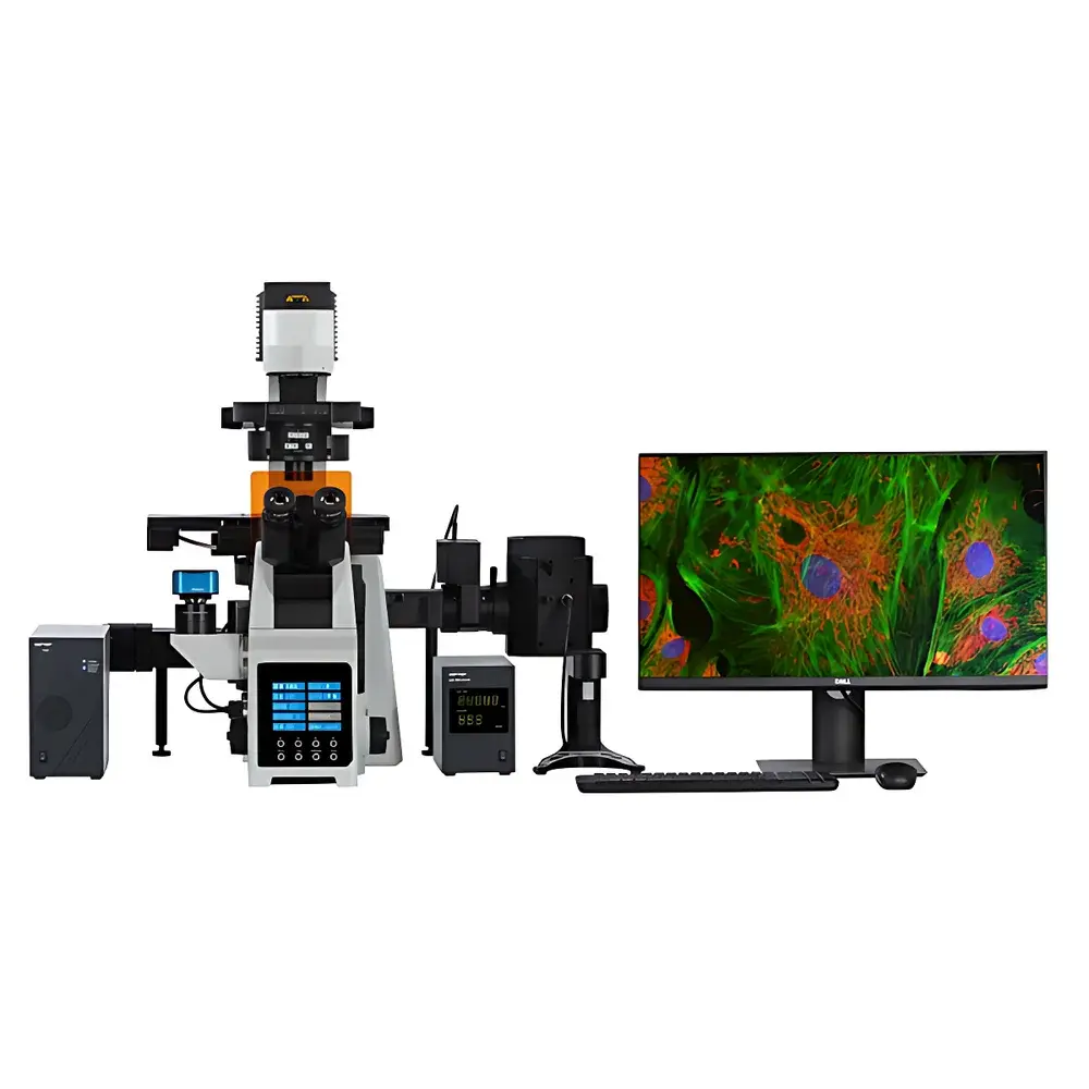
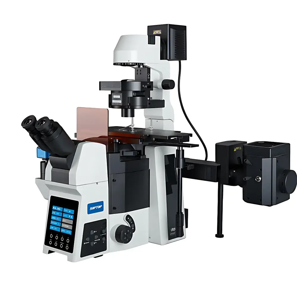
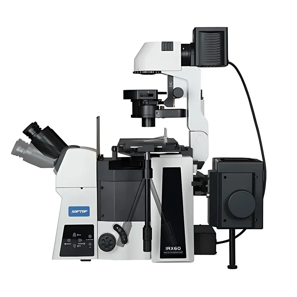
The SOPTOP IRX60 is a fully motorized, research-grade inverted fluorescence microscope engineered for high-fidelity live-cell imaging, electrophysiology (e.g., patch-clamp), microinjection, and long-term time-lapse observation. Its optical architecture is based on an infinity-corrected, modular beam path design optimized for simultaneous multi-modal contrast—fluorescence, phase contrast, differential interference contrast (DIC), and brightfield—with minimal mechanical coupling between subsystems. The system employs a high-stability mechanical platform, thermally stabilized mercury excitation source, and precisely aligned Köhler illumination pathways to ensure photometric consistency across modalities and sessions. Designed for rigorous laboratory environments, the IRX60 meets core requirements of GLP-compliant workflows and supports audit-ready documentation when integrated with validated software platforms.
Key Features
- Fully Motorized Optomechanical Control: All critical components—including objective turret, fluorescence filter wheel, ND filter wheel, condenser aperture, focus drive, and electronic shutter—are driven by precision stepper motors with closed-loop positional feedback, enabling repeatable, scriptable operation.
- Ergonomic Tilting Observation Head: Adjustable inclination from 20° to 45° allows optimal eye-point positioning up to 78 mm above stage level (at 65 mm IPD), eliminating need for height-adjustable chairs or ocular lifters—critical for extended operator sessions and standing-mode workflows.
- Modular Multi-Contrast Condenser System: A motorized 7-position condenser (NA ≥ 0.55, WD ≥ 27 mm) accommodates interchangeable annuli and prisms for phase contrast (φ30 mm) and DIC (φ38 mm), with integrated polarizer assembly for simplified polarization-based assays.
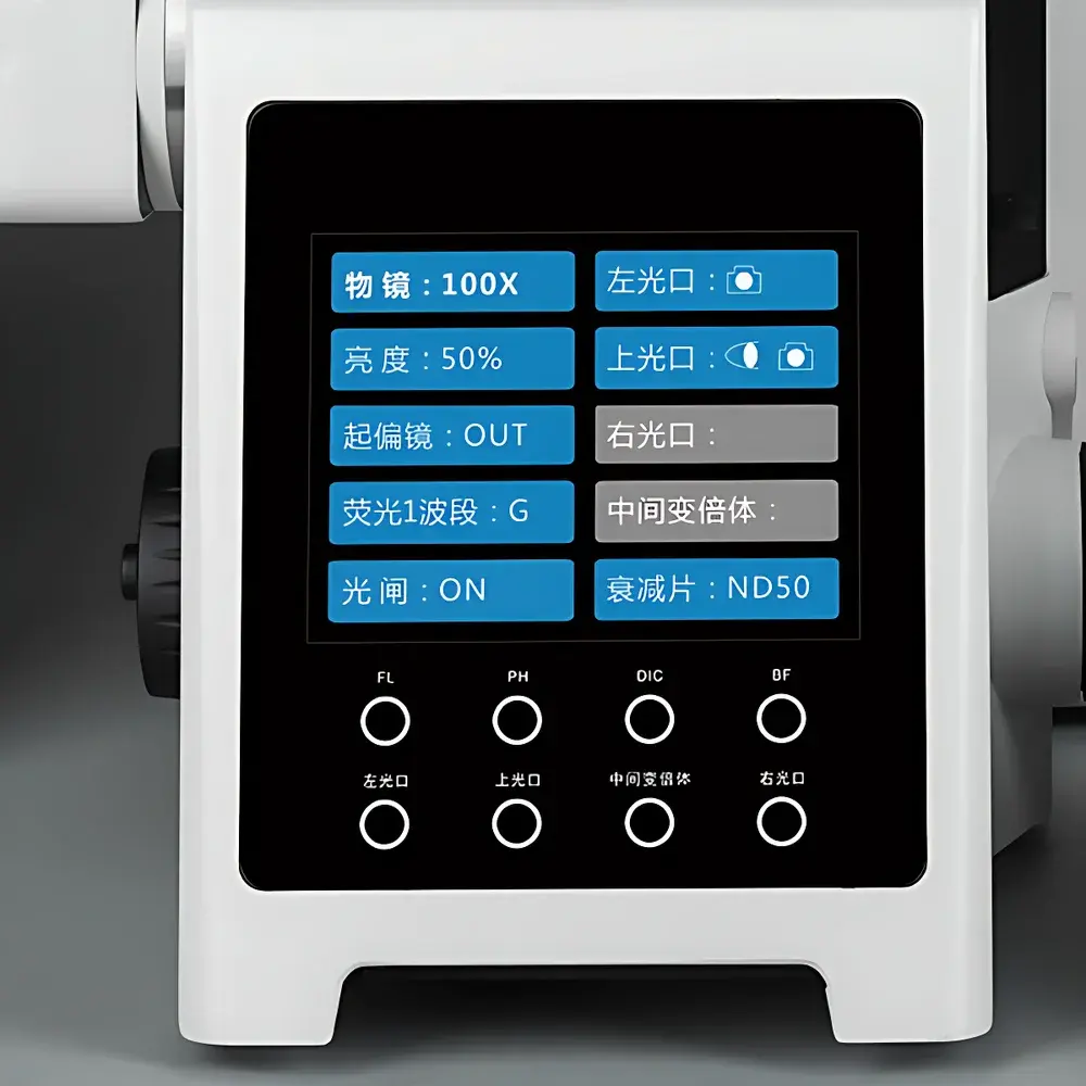
- Integrated Front-Panel Digital Interface: A high-resolution OLED display shows real-time status of objective magnification, transmitted light intensity, active fluorescence band, shutter state, and filter positions—enabling rapid configuration without software dependency.
- Spill-Resistant Optical Architecture: Drain grooves beneath the objective nosepiece channel accidental media overflow away from optical surfaces and electrical modules, significantly reducing contamination risk during cell culture handling.
- Expandable Dual-Light-Path Design: Supports up to 16 fluorescence filter sets via optional dual-layer filter cube holders; multiple side and rear ports accommodate simultaneous attachment of sCMOS cameras, confocal scanning units, infrared detectors, or laser coupling optics.
Sample Compatibility & Compliance
The IRX60 is optimized for standard tissue culture formats—including 35 mm, 60 mm, and 100 mm Petri dishes, multi-well plates (6–96-well), and chambered coverslips—with full compatibility for glass-bottom vessels (0.13–0.17 mm thickness). Its long-working-distance semi-apochromatic and super-apochromatic objectives incorporate correction collars to compensate for spherical aberration induced by variable cover-glass thicknesses and immersion media refractive indices. While not certified as a medical device under FDA 21 CFR Part 820 or EU MDR, the IRX60 conforms to IEC 61000-6-3 (EMC emissions) and IEC 61000-6-2 (immunity) standards. Its motorized control architecture supports integration with systems compliant with FDA 21 CFR Part 11 when paired with validated third-party acquisition software featuring electronic signatures and audit trails.
Software & Data Management
The IRX60 operates natively with SOPTOP’s proprietary imaging suite, which provides full hardware abstraction for automated acquisition protocols—including multi-channel fluorescence time series, Z-stack acquisition with hardware-triggered autofocus, extended depth-of-field synthesis, tile-scan mosaicking, and quantitative intensity profiling. The software implements ISO/IEC 17025-aligned metadata tagging (including exposure time, gain, binning, objective ID, filter set, and environmental timestamps) and exports TIFF, OME-TIFF, and HDF5 formats. Exported datasets retain embedded calibration parameters for downstream analysis in ImageJ/Fiji, MATLAB, or Python-based pipelines (e.g., Napari, CellProfiler). Optional SDK support enables custom scripting via C++ or Python APIs for integration into automated screening platforms.
Applications
- Live-Cell Dynamics: Long-duration tracking of subcellular organelles, cytoskeletal remodeling, and vesicle trafficking using low-phototoxicity fluorescence protocols.
- Electrophysiology Support: Precise alignment and stable visualization during whole-cell patch-clamp recordings, facilitated by vibration-damped stage and zero-drift focus stabilization.
- Microinjection & Embryology: High-contrast DIC and phase contrast enable clear visualization of pronuclei, spindle apparatus, and blastomeres in zygotes and early embryos.
- Co-Culture & 3D Spheroid Imaging: Extended working distance objectives and adjustable condenser clearance permit unobstructed access for micromanipulators and perfusion tubing in complex assay setups.
- Multi-Modal Phenotyping: Simultaneous acquisition of structural (phase/DIC), functional (Ca²⁺ indicators), and morphological (DAPI/Hoechst) signals across defined ROI grids for high-content analysis.
FAQ
Is the IRX60 compatible with third-party cameras and controllers?
Yes—the system provides TTL, USB 3.0, and RS-232 interfaces for synchronization with external sCMOS/EMCCD cameras, piezo Z-drives, and environmental chambers.
Does the IRX60 support oil immersion objectives?
Yes—select infinity-corrected apochromatic objectives (e.g., 60X/100X NA 1.40) are designed for oil immersion and include correction collars for optimal performance across cover-glass thicknesses.
Can the motorized stage be upgraded post-purchase?
Yes—manual stages are field-upgradable to motorized XY stages via factory-certified retrofit kits with calibrated encoders and firmware integration.
What fluorescence filter sets are included by default?
Standard delivery includes B1 (FITC), G1 (TRITC), and UV1 (DAPI) filter cubes; additional sets (e.g., Cy5, mCherry, GFP/mCherry dual-band) are available as configurable options.
Is remote operation supported?
Yes—TCP/IP-based network control enables full instrument operation via LAN-connected PCs or secure cloud-accessible gateways, supporting centralized lab management and multi-user scheduling.
Related Products


