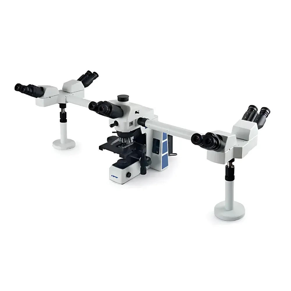
SOPTOP RX50DO5 Multi-Observer Upright Biological Microscope
| Brand | SOPTOP |
|---|---|
| Origin | Zhejiang, China |
| Manufacturer Type | Direct Manufacturer |
| Country of Origin | China |
| Model | RX50DO5 |
| Instrument Type | Upright Microscope |
| Eyepiece Configuration | Trinocular |
| Price Range | USD 16,800 – 28,000 (FOB China) |
| Compliance | CE-marked per EN 61000-6-3/6-4 & EN 61000-6-2 |
Overview
The SOPTOP RX50DO5 is a purpose-engineered trinocular upright biological microscope optimized for collaborative observation in academic, clinical, and research environments. Built upon Köhler illumination principles and utilizing an infinity-corrected optical path (180 mm tube length), the system delivers high-contrast, color-accurate imaging across wide-field eyepieces (WF10×/22 mm standard). Its core architecture supports simultaneous real-time viewing by up to five observers—two via lateral auxiliary tubes, two via integrated side-monitor ports, and one via the primary trinocular phototube—without optical splitting losses or image degradation. Unlike conventional shared-view solutions relying on beam splitters or digital relay, the RX50DO5 employs a mechanically synchronized dual-path light distribution system that preserves native resolution and brightness at each viewing station. This design eliminates parallax-induced misalignment during group analysis of histological sections, live-cell cultures, or hematological smears—critical for consensus-based diagnosis in pathology training or peer-reviewed morphological assessment in parasitology and oncology labs.
Key Features
- Trinocular head with 30° inclined binocular viewing tubes and dedicated C-mount phototube for camera integration
- Dual lateral observation ports (2×) enabling direct optical viewing by two additional users without digital latency or resolution compromise
- Large-format mechanical stage (187 × 168 mm) with bi-directional linear rail movement, calibrated X-Y vernier scales (0.1 mm resolution), and ergonomic retractable dual-hand control levers
- LED-transmitted illumination system (6 V / 30 W equivalent, 50,000-hour lifetime) with adjustable intensity, centerable condenser (NA 1.25, swing-out top lens), and built-in field diaphragm
- Skin-friendly polymer-coated chassis with anti-static surface treatment (surface resistivity < 10⁹ Ω/sq) and low-thermal-conductivity housing to minimize operator thermal fatigue during extended sessions
- Integrated LED pointer module synchronized with primary optical axis—visible in all viewing paths including side monitors—enabling precise annotation during instruction or case review
Sample Compatibility & Compliance
The RX50DO5 accommodates standard glass slides (26 × 76 mm), Petri dishes (up to 100 mm diameter), and multi-well plates (6–96-well) using optional stage adapters. It supports brightfield, phase contrast (with optional PH condenser and objectives), and optional polarized light configurations. All optical components comply with ISO 8578 (microscope objective labeling standards) and are certified for use under GLP-compliant workflows when paired with validated digital imaging systems. The instrument meets electromagnetic compatibility requirements per EN 61000-6-3 (emission) and EN 61000-6-2 (immunity), and electrical safety conforms to IEC 61010-1:2010 + A1:2019. While not FDA-cleared as a medical device, its optical performance aligns with ASTM E2877-22 (Standard Guide for Microscope Calibration) and USP recommendations for microscopy-based pharmaceutical quality control.
Software & Data Management
The RX50DO5 interfaces natively with SOPTOP’s proprietary ToupView 5.x software suite (Windows/macOS/Linux compatible) via USB 3.0, supporting real-time streaming at 30 fps (1920 × 1080), time-lapse acquisition, and measurement annotation with pixel calibration traceability. When connected to external displays or projectors via HDMI output from the included imaging module, the system enables synchronous multi-display presentation—ideal for classroom instruction or multidisciplinary tumor board reviews. Audit trail functionality (user login, timestamped capture logs, metadata embedding) satisfies basic 21 CFR Part 11 readiness when deployed with network-authenticated file storage. Raw image data is saved in TIFF/RAW formats with embedded EXIF metadata (objective magnification, illumination intensity, exposure settings).
Applications
This platform is routinely deployed in university histology teaching laboratories for comparative tissue morphology instruction; in hospital pathology departments for resident-led slide seminars requiring concurrent expert annotation; in parasitology field stations for rapid malaria or filariasis smear evaluation by multi-tier diagnostic teams; and in immunology core facilities for collaborative scoring of IHC-stained tumor sections. Its mechanical stability and vibration-damped base also support long-duration live-cell imaging when combined with environmental chamber accessories (not included). The large stage and lateral port geometry facilitate hands-on manipulation of specimens during demonstration—e.g., microinjection setup verification or micromanipulator alignment checks.
FAQ
Can the RX50DO5 be integrated into existing LIMS or hospital PACS networks?
Yes—when used with SOPTOP’s DICOM-compliant export module (optional license), captured images and annotated reports can be transmitted directly to PACS via DICOM SCU protocol or exported as HL7-compatible structured reports.
Is phase contrast capability built-in or requires add-on components?
Phase contrast functionality requires optional PH-series condenser and matched phase objectives (e.g., PH10×, PH40×); these are available as factory-configured kits but not included in base pricing.
What is the maximum working distance supported for objective lenses?
The standard objective turret accepts lenses with working distances up to 10.6 mm (e.g., SOPTOP PLN40×/0.65 WD=0.66 mm; PLN100×/1.25 oil WD=0.13 mm); custom long-working-distance objectives (e.g., 20 mm WD) may be mounted with adapter rings.
Does the LED pointer remain visible under all contrast modes?
The coaxial LED pointer is optically coupled to the main light path and remains fully visible in brightfield, phase contrast, and polarized light—though contrast may vary slightly depending on background luminance and condenser aperture setting.
Related Products


