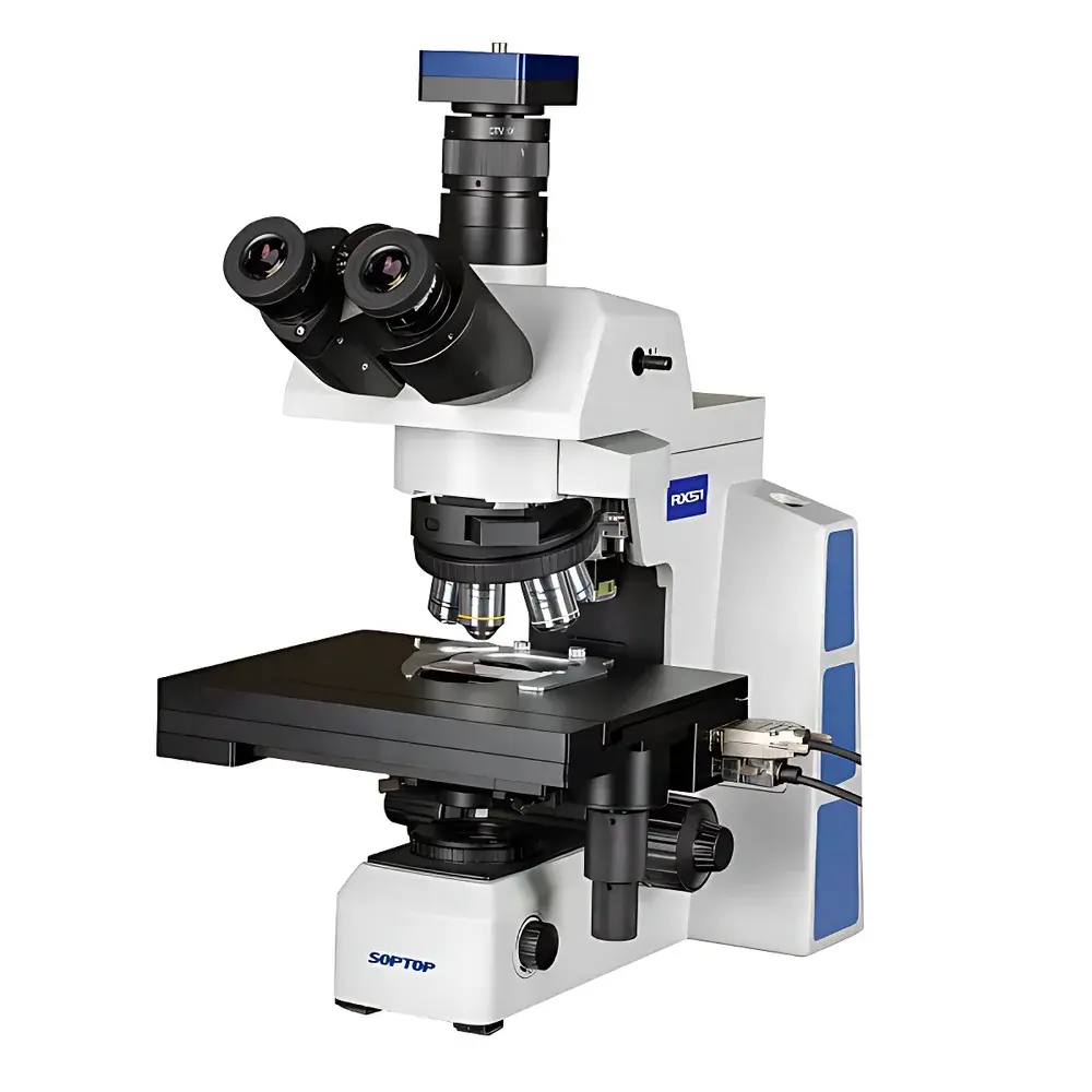
SOPTOP RX51 Upright Biological Microscope
| Brand | SOPTOP |
|---|---|
| Origin | Zhejiang, China |
| Manufacturer Type | Original Equipment Manufacturer (OEM) & ODM |
| Country of Origin | China |
| Model | RX51 |
| Instrument Type | Upright Microscope |
| Head Configuration | Trinocular |
| Illumination | LED-based Köhler illumination with homogenized field optics |
| Focus Drive | Motorized XYZ stage with manual override |
| Imaging Mode | Real-time digital capture, automated whole-slide scanning, multi-layer Z-stack acquisition |
| Software Features | Auto-focus, auto-white balance, exposure/gain control, panoramic stitching, GLP-compliant metadata logging |
Overview
The SOPTOP RX51 Upright Biological Microscope is an integrated optical platform engineered for high-fidelity brightfield and phase contrast microscopy in life science laboratories, clinical pathology departments, and academic research facilities. Built upon a rigid mechanical chassis and optimized optical path design, the RX51 employs infinity-corrected optical systems compatible with standard 4×–100× objective lenses (including oil immersion). Its core architecture supports both conventional visual observation via trinocular head—diverting ~20% light to eyepieces and ~80% to the imaging port—and fully automated digital workflows. Unlike legacy upright microscopes limited to static imaging, the RX51 integrates motorized XYZ precision mechanics with synchronized camera control to enable repeatable, operator-independent slide digitization. This dual-mode capability—manual exploration and programmable scanning—aligns with evolving demands for traceable, auditable, and scalable microscopy data generation in regulated environments.
Key Features
- Trinocular optical head with adjustable interpupillary distance and diopter compensation, supporting simultaneous ocular viewing and high-resolution C-mount or F-mount camera integration.
- Motorized XYZ translation stage (X/Y: ±50 mm travel; Z: 25 mm fine focus range) with positional repeatability ≤1 µm and programmable speed profiles for seamless scanning trajectory execution.
- Köhler illumination system enhanced by fly-eye lens array, ensuring uniform intensity distribution across the entire field of view (FOV) at all magnifications—from 40× overview to 1000× oil-immersion resolution—without hot spots or edge falloff.
- Long-life, color-stable 6,000 K white LED illumination (≥50,000 h rated lifetime), compliant with IEC 62471 Photobiological Safety standards, eliminating UV hazard and thermal drift during extended acquisition sessions.
- Dedicated RX51 Control Suite software providing FDA 21 CFR Part 11–ready audit trail logging, user role-based access control, and timestamped metadata embedding (objective ID, exposure time, gain, stage coordinates, calibration status).
Sample Compatibility & Compliance
The RX51 accommodates standard 1″ × 3″ (25 × 75 mm) glass microscope slides with thicknesses of 0.9–1.2 mm, including stained histology sections (H&E, IHC), cytology preparations, live-cell culture dishes (35 mm petri dishes with #1.5 coverslip bottom), and whole-mount tissue specimens up to 2 mm in height. Stage clips and optional mechanical specimen holders ensure secure positioning during automated scans. The system meets ISO 10993-5 biocompatibility requirements for components contacting sample carriers and adheres to CE marking directives for laboratory equipment (2014/30/EU EMC and 2014/35/EU LVD). All firmware and software modules are validated per GLP and GMP Annex 11 principles for data integrity, including electronic signature support and change-controlled version management.
Software & Data Management
RX51 Control Suite is a Windows-based application built on modular architecture to decouple acquisition, processing, and archival functions. It supports real-time preview at full sensor resolution (up to 20 MP), dynamic exposure adjustment (0.1–5000 ms range), analog/digital gain scaling (0–24 dB), and hardware-triggered synchronization with external devices (e.g., environmental chambers or perfusion pumps). Automated scanning routines include single-layer panoramic tiling (with overlap-based seam correction), multi-focus Z-stack acquisition (user-defined slice intervals from 0.2–10 µm), and region-of-interest (ROI) targeted scanning. Export formats include TIFF (uncompressed), JPEG2000 (lossless compression), and OME-TIFF (Open Microscopy Environment compliant with Bio-Formats ingestion). Raw image datasets retain embedded EXIF and custom metadata fields required for ISO/IEC 17025 accreditation documentation.
Applications
The RX51 serves as a primary imaging instrument in university core facilities for undergraduate histology instruction and graduate-level cell biology research, particularly where longitudinal tracking of morphological changes in fixed or live specimens is required. In clinical pathology labs, it functions as a pre-scanner for digital pathology validation—generating reference-quality images prior to deployment on high-throughput whole-slide scanners. Its robust mechanical design and calibrated optical train also support QC/QA tasks in pharmaceutical manufacturing, including particulate analysis in injectables (per USP ) and sterility test membrane examination (USP ). Additionally, the system supports fluorescence module retrofitting (optional LED excitation cubes), extending utility into immunofluorescence co-localization studies under controlled ambient light conditions.
FAQ
Is the RX51 compatible with third-party cameras and software platforms?
Yes—the microscope features standard C-mount and F-mount interfaces, and its SDK supports TWAIN and GenICam protocols for integration with commercial image analysis packages (e.g., HALO, Visiopharm, ImageJ/Fiji via plugin architecture).
Does the motorized stage support closed-loop feedback for positional accuracy?
The XYZ stage utilizes stepper motors with optical encoders, delivering open-loop positional accuracy of ±1.5 µm and repeatability of ≤1 µm—validated per ISO 9283 standards for industrial manipulators.
Can the RX51 meet regulatory requirements for clinical diagnostic use?
While the base system is Class I medical device compliant (CE-marked), diagnostic interpretation requires additional validation per IVD Directive 2017/746 (IVDR) and local health authority approval; SOPTOP provides technical documentation packages to support such submissions.
What maintenance protocols are recommended for long-term optical performance?
Annual calibration of illumination uniformity and focus linearity is advised using NIST-traceable reference slides; cleaning follows ISO 10110-7 standards for coated optical surfaces, with manufacturer-supplied lens tissues and solvent-free wipes.
Is remote operation supported over network infrastructure?
Yes—RX51 Control Suite includes secure RDP-compatible remote desktop mode and RESTful API endpoints for integration into LIMS or ELN systems, enabling centralized instrument monitoring and batch job queuing across multi-site deployments.
Related Products


