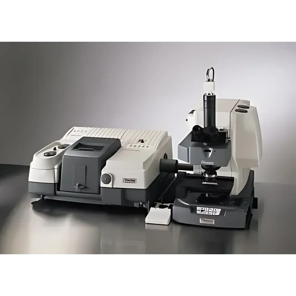
Thermo Fisher Scientific Continuum XL Infrared Imaging Microscope
| Brand | Thermo Fisher Scientific |
|---|---|
| Origin | USA |
| Manufacturer Type | Original Equipment Manufacturer (OEM) |
| Import Status | Imported |
| Model | Continuum XL |
| Pricing | Available upon request |
Overview
The Thermo Fisher Scientific Continuum XL Infrared Imaging Microscope is a high-performance, research-grade FTIR microspectroscopy platform engineered for spatially resolved chemical characterization at the microscopic level. Built upon a fully motorized optical bench and integrated with advanced interferometry, the system operates across the mid-infrared (MIR) and near-infrared (NIR) spectral ranges (typically 7500–600 cm⁻¹, extendable with optional detectors). Its core measurement principle relies on Fourier-transform infrared spectroscopy coupled with precise sample positioning and focal-plane array (FPA) or linear array detection—enabling rapid, label-free molecular mapping of heterogeneous samples without destructive preparation. Unlike conventional single-point IR microscopes, the Continuum XL supports scalable acquisition architectures: users may configure the system for traditional point-spectral analysis, dual-linear array imaging, or high-throughput FPA-based hyperspectral imaging—making it suitable for both targeted microanalysis and comprehensive chemical distribution studies in materials science, pharmaceuticals, forensics, and life sciences.
Key Features
- Modular architecture supporting seamless transition from single-point microspectroscopy to dual-linear array imaging and FPA-based infrared hyperspectral imaging
- Patented optical design inherited from the Continuum series—including automated beam alignment, dynamic aperture control, and vibration-damped optical path
- Software-selectable single- or dual-aperture mode: optimized for either high-spatial-resolution point analysis (down to ~3–5 µm depending on wavelength and objective) or wide-field chemical imaging
- Multi-modal sampling compatibility: transmission, reflection, grazing-angle reflection, and attenuated total reflection (ATR) with interchangeable objectives and accessories
- Five detector options available—including DTGS, MCT (single- and dual-element), and liquid-nitrogen-cooled FPA arrays—to match spectral range, sensitivity, and speed requirements
- Dual-linear array detector configuration enabling parallel data acquisition across two adjacent rows of pixels, improving frame rate and duty cycle without sacrificing spatial fidelity
- Motorized XYZ stage with three-speed preview mode (coarse/medium/fine) for rapid region-of-interest localization and reproducible positioning within ±0.5 µm repeatability
- High-fidelity visible-light imaging subsystem (10 MP CMOS) co-registered with IR data for precise morphological correlation
- USB 2.0 interface for real-time spectral streaming and synchronized hardware control; compatible with optional Ethernet-based remote operation
Sample Compatibility & Compliance
The Continuum XL accommodates solid, thin-film, particulate, and semi-transparent biological specimens—including polymer blends, pharmaceutical tablets, tissue sections, microplastics, and forensic trace evidence. Sample mounting follows standard IR microscopy protocols (e.g., KBr pellet compression, diamond anvil cells, reflective substrates). The system complies with ISO 17025 requirements for analytical instrument qualification and supports GLP/GMP workflows through audit-trail-enabled software (OMNIC Paradigm). Optional validation packages include IQ/OQ documentation aligned with ASTM E1252 (Standard Practice for General Techniques for Infrared Microanalysis) and USP (Infrared Absorption Spectrophotometry). Data integrity meets FDA 21 CFR Part 11 criteria when deployed with appropriate electronic signature and access-control configurations.
Software & Data Management
Controlled by Thermo Fisher’s OMNIC Paradigm software, the Continuum XL provides unified acquisition, processing, and visualization for both spectral and imaging datasets. The interface supports real-time spectral preview, automated background correction, multivariate analysis (PCA, cluster analysis, classical least-squares), and pixel-by-pixel spectral library searching (Spectral ID). All raw interferograms and processed spectra are stored in vendor-neutral HDF5 format with embedded metadata (instrument parameters, stage coordinates, detector settings). Batch processing pipelines enable automated QA/QC reporting, while export modules support CSV, JCAMP-DX, and ENVI-compatible formats for third-party chemometric tools. Software updates follow a controlled release cycle compliant with IEC 62304 for medical device software lifecycle management.
Applications
- Pharmaceutical: Active pharmaceutical ingredient (API) distribution mapping in tablets, polymorph identification in crystalline domains, coating uniformity assessment
- Materials Science: Phase segregation analysis in polymer composites, oxidation state mapping in battery electrodes, contaminant identification in semiconductor wafers
- Life Sciences: Lipid/protein distribution in frozen-hydrated tissue sections, disease biomarker localization in histopathology, cell membrane composition profiling
- Forensics: Fiber dye analysis, paint layer stratigraphy, gunshot residue particle discrimination
- Environmental: Microplastic polymer typing and weathering state assessment, soil organic matter functional group mapping
FAQ
What spectral range does the Continuum XL support?
The base configuration covers 4000–600 cm⁻¹ (mid-IR); optional NIR extensions reach up to 7500 cm⁻¹, and deep-IR options extend down to 550 cm⁻¹ using specialized beamsplitters and detectors.
Can the system perform ATR imaging?
Yes—when equipped with a motorized ATR objective (e.g., 16× germanium crystal), the Continuum XL enables contact-mode chemical imaging with spatial resolution limited primarily by crystal geometry and pressure uniformity.
Is FPA imaging supported natively?
Yes—the system accepts 64×64 and 128×128 mercury cadmium telluride (MCT) FPA detectors, with full hardware synchronization, dead-pixel correction, and non-uniformity compensation algorithms embedded in OMNIC Paradigm.
How is calibration maintained across imaging modes?
Wavelength accuracy is verified daily via built-in polystyrene reference film; spatial calibration is performed using NIST-traceable stage encoders and certified grating standards, with automated recalibration triggered after objective changes or thermal drift compensation cycles.
Does the system integrate with laboratory information management systems (LIMS)?
Via optional API modules, OMNIC Paradigm supports HL7 and ASTM E1714-compliant data exchange protocols, enabling direct spectral metadata ingestion into enterprise LIMS or ELN platforms.


