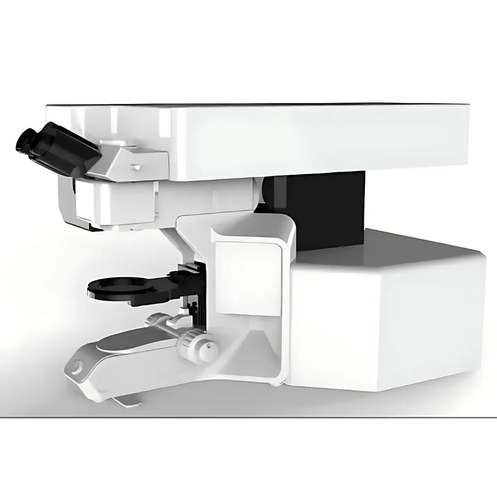
VibroniX LDM Laser Microdissection System
| Brand | VibroniX |
|---|---|
| Origin | Jiangsu, China |
| Manufacturer Type | Authorized Distributor |
| Country of Origin | China |
| Model | LDM |
| Pricing | Available Upon Request |
Overview
The VibroniX LDM Laser Microdissection System is a precision optical instrument engineered for contact-free, high-resolution isolation of specific cellular or tissue regions directly under microscopic visualization. Operating on the principle of focused near-infrared or UV laser ablation—typically at 355 nm (UV) or 1064 nm (NIR)—the system enables spatially selective dissection with micron-level accuracy. Unlike mechanical microdissection or manual scraping methods, the LDM employs a galvanometer-driven scanning mirror system to steer the laser beam across the sample plane without moving the specimen stage or objective. This optical beam steering architecture ensures minimal thermal diffusion, preserves molecular integrity (including RNA and protein), and maintains sample stability throughout the procedure—critical for downstream genomic, proteomic, or metabolomic analysis.
Key Features
- Optical Beam Steering Architecture: A high-speed galvanometric mirror system directs the laser beam with sub-micron positional repeatability, enabling real-time, dynamic cutting paths while the sample remains stationary—eliminating stage vibration artifacts and improving reproducibility.
- Gravity-Based Collection Mechanism: Dissected tissue fragments are released by controlled laser ablation and collected passively via gravity into standard microcentrifuge tubes or PCR plates. This non-contact, force-free method prevents mechanical stress, avoids cross-contamination, and supports collection of heterogeneous or fragile structures—including single cells, neural dendrites, or stromal layers—without size or geometry constraints.
- Adjustable High-Power Laser Source: Integrated diode-pumped solid-state (DPSS) laser with continuously tunable power (1–100 mW), adjustable spot size (1–15 µm), variable pulse frequency (1–50 kHz), and programmable scanning velocity (0.1–5 mm/s). Focus is motorized and software-synchronized for depth-resolved sectioning in thick samples up to 100 µm.
- Ergonomic Upright Microscope Platform: Equipped with a research-grade upright microscope featuring infinity-corrected optics, motorized Z-focus, high-NA objectives (10×–63×), and uniform LED illumination (5500 K color temperature) optimized for brightfield, phase contrast, and fluorescence pre-screening—ensuring optimal morphological assessment prior to dissection.
- Workflow-Centric Software Suite: Intuitive, icon-driven interface supporting annotation-based region-of-interest (ROI) definition, multi-layer mask management, batch processing, and export of coordinate metadata in standardized formats (e.g., .xml, .csv). Includes built-in calibration routines compliant with ISO/IEC 17025 traceability requirements.
Sample Compatibility & Compliance
The LDM system accommodates a broad range of fixed and unfixed biological specimens, including cryosections (4–20 µm), paraffin-embedded sections (deparaffinized), live-cell monolayers (on glass-bottom dishes), and whole-mount tissues (up to 2 mm thickness with optional water-dipping objectives). It supports standard histopathology substrates (e.g., PEN membrane slides, Zeiss MembraneSlides) and integrates seamlessly with common nucleic acid extraction kits (Qiagen, Arcturus). The system meets essential regulatory expectations for analytical instrumentation used in GLP-compliant laboratories: audit trail logging (user ID, timestamp, parameter changes), electronic signature support per FDA 21 CFR Part 11, and alignment with ISO 13485 quality management principles for IVD-adjacent applications.
Software & Data Management
The proprietary LDM Control Software v3.2 provides full instrument control, image overlay registration (live camera feed + imported TIFF/SVS), ROI segmentation using threshold-based or AI-assisted contour detection (optional plugin), and automated laser path generation. All acquisition parameters—including laser energy density (J/cm²), dwell time, and z-stack offsets—are stored with each dataset in a relational SQLite database. Export options include FAIR-compliant metadata (MIAME/MIAPE extensions), DICOM-SR for pathology integration, and direct API linkage to LIMS platforms via RESTful endpoints. Raw image data retains EXIF-like embedded calibration tags for retrospective validation.
Applications
- Genomic profiling of tumor subclones from heterogeneous FFPE sections
- Single-cell RNA-seq preparation from defined brain nuclei or pancreatic islets
- Isolation of endothelial cells from vascularized tissue explants for ex vivo angiogenesis assays
- Precision excision of fungal hyphae from plant root cross-sections for host-pathogen transcriptomics
- Targeted removal of pigment granules in retinal pigment epithelium (RPE) for oxidative stress biomarker studies
- Quality control in biobanking—verification and retrieval of annotated archival tissue cores
FAQ
What laser wavelength options are available on the LDM system?
Standard configuration includes a 355 nm UV DPSS laser; optional 1064 nm NIR module is available for deeper penetration in scattering tissues.
Can the LDM be integrated with third-party fluorescence imaging systems?
Yes—via TTL-triggered synchronization and external camera input (HDMI/USB3), enabling multimodal correlative workflows (e.g., confocal-guided microdissection).
Is the software validated for regulated environments (e.g., clinical diagnostics)?
While not FDA-cleared as an IVD device, the software architecture supports 21 CFR Part 11 compliance when deployed with enterprise authentication and network-attached storage.
What is the minimum feature size resolvable and dissectable?
With 63× oil immersion and 355 nm laser, theoretical diffraction-limited resolution is ~220 nm; practical dissection precision is ≤1 µm for contiguous features.
Does the system require special environmental conditions (e.g., vibration isolation)?
No active damping is required; however, operation on a standard optical table (or equivalent rigid surface) is recommended to maintain long-term galvo alignment stability.


