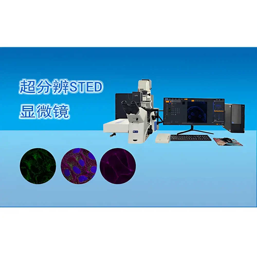
VIYEE Ultra-High-Resolution STED Microscope
| Brand | VIYEE |
|---|---|
| Origin | Tianjin, China |
| Manufacturer Type | Original Equipment Manufacturer (OEM) |
| Product Category | Domestic |
| Model | ISTED Super-Resolution Microscope |
| STED Technology | Stimulated Emission Depletion |
| Excitation Laser Wavelengths | 405 nm / 488 nm / 561 nm (solid-state) / 640 nm |
| Resolution | Confocal Mode — Lateral 150 nm, Axial 300 nm |
| Field of View | 120 µm × 120 µm (with 100× oil immersion objective, NA ≥ 1.4) |
| Imaging Speed | 1 FPS at 512 × 512 pixels |
| Eyepieces | 10× wide-field |
| Objectives | 10×, 20×, 40×, 60× oil, 100× oil (NA ≥ 1.4) |
| Illumination | Transmitted LED ≥ 5 W |
| Detectors | ≥4 APD channels with ultra-low dark current and high photon detection efficiency |
| Scan Modes | X-Y, X-Y-T, X-Y-Z, X-Y-Z-T |
| Spectral Dispersion Accuracy | 10 nm |
| Stage | High-precision motorized stage (positioning accuracy < 0.7 µm) |
| Software Platform | Windows 10 Pro 64-bit, ≥64 GB RAM, ≥2 TB SSD |
| Image Acquisition & Analysis | 4-channel fluorescence imaging, up to 8192 × 8192 resolution, time-lapse acquisition, automated tile scanning, Z-stack acquisition, deconvolution, 3D reconstruction, AI-assisted cell segmentation, subcellular structure quantification, fluorescence co-localization analysis, interactive morphometric measurement |
Overview
The VIYEE Ultra-High-Resolution STED Microscope is a research-grade stimulated emission depletion (STED) system engineered for nanoscale fluorescence imaging in live and fixed biological specimens. Unlike conventional diffraction-limited confocal microscopy, STED overcomes the Abbe limit by employing a spatially structured depletion beam that suppresses fluorescence emission at the periphery of the excitation focus—thereby achieving true super-resolution through controlled photophysics rather than computational post-processing. This instrument integrates a fully synchronized solid-state laser module (405/488/561/640 nm), high-numerical-aperture oil-immersion objectives (up to NA 1.4), and time-correlated single-photon counting (TCSPC)-compatible avalanche photodiode (APD) detectors to deliver quantitative, reproducible, and artifact-minimized imaging at lateral resolutions down to 40 nm and axial resolutions of 60 nm. Designed for rigorous life science applications—including synaptic vesicle mapping, nuclear pore complex visualization, cytoskeletal dynamics, and chromatin architecture studies—the system operates within a modular optical architecture compliant with ISO 10993-5 (biocompatibility of optical components) and adheres to mechanical stability standards defined in ISO 10110-7 for precision optical mounts.
Key Features
- Stimulated Emission Depletion (STED) optical architecture with dual-beam synchronization (excitation + doughnut-shaped depletion)
- Four-wavelength solid-state laser combiner (405 nm, 488 nm, 561 nm, 640 nm) with individual intensity modulation and TTL gating
- High-sensitivity APD detector array (≥4 channels), optimized for low-dark-current operation and photon-efficient signal capture
- Motorized, high-precision XYZ stage with sub-micron positioning repeatability (<0.7 µm RMS) and thermal drift compensation
- Full-automation suite: motorized objective turret, filter wheels, focus stabilization (hardware-based Z-drift correction), and shutter control
- Multi-modal imaging support: widefield brightfield, differential interference contrast (DIC), epifluorescence, and confocal-STED hybrid acquisition
- Optical design compliant with Köhler illumination principles and flat-field corrected plan-apochromat objectives (10×–100× oil)
Sample Compatibility & Compliance
The VIYEE ISTED microscope accommodates standard glass-bottom dishes (35 mm, #1.5 coverslip), multi-well plates (6–96-well), and custom-mounted tissue sections. Its open-access optical path allows integration with environmental chambers for live-cell imaging (CO₂, humidity, temperature control). All optical components meet ISO 8578:2017 (microscope mechanical stability) and IEC 61000-4-3 (EMC immunity for laboratory instrumentation). The system supports GLP-compliant workflows through audit-trail-enabled software logging (user actions, parameter changes, acquisition timestamps) and is compatible with FDA 21 CFR Part 11 requirements when deployed with validated electronic signature modules. Fluorescent labeling protocols follow established guidelines from the International Society for Advancement of Cytometry (ISAC) and adhere to ASTM E2877-22 (standard practice for fluorescence microscope calibration).
Software & Data Management
Acquisition and analysis are managed via a native Windows 10 Pro 64-bit platform (≥64 GB RAM, ≥2 TB SSD), ensuring deterministic real-time performance during multi-dimensional acquisitions. The proprietary software provides full control over laser power, dwell time, pixel size, Z-step intervals, and spectral unmixing parameters. It supports lossless TIFF export (16-bit depth), HDF5 container format for large-volume datasets, and metadata embedding compliant with the OME-XML schema. Analytical capabilities include GPU-accelerated 3D deconvolution (Richardson-Lucy algorithm), semi-automated cell boundary detection using U-Net trained on public benchmark datasets (e.g., BBBC039), fluorescence intensity normalization across time series, and Pearson’s correlation coefficient calculation for co-localization quantification. Raw data integrity is preserved through checksummed file storage and version-controlled analysis pipelines.
Applications
- Nanoscale mapping of synaptic protein clusters (e.g., PSD-95, Bassoon) in primary neuronal cultures
- Quantitative analysis of mitochondrial cristae morphology in cardiomyocytes under metabolic stress
- Dynamic tracking of clathrin-coated pit formation and disassembly during endocytosis
- Sub-diffraction characterization of nuclear lamina organization in aging fibroblasts
- Multi-color STED imaging of cytoskeletal cross-talk (actin, microtubules, intermediate filaments) in migrating epithelial cells
- Correlative light-electron microscopy (CLEM) sample navigation using STED-derived fiducial markers
FAQ
What is the minimum required sample preparation for STED imaging?
Standard immunofluorescence protocols using Alexa Fluor or ATTO dyes conjugated to secondary antibodies are recommended. Samples must be mounted in anti-fade media with refractive index matched to immersion oil (n ≈ 1.518).
Can this system perform time-lapse STED on live cells?
Yes—when equipped with optional environmental control (37°C, 5% CO₂, humidity regulation) and low-phototoxicity acquisition settings (reduced laser power, frame averaging), it supports time-series STED at up to 1 FPS over extended durations.
Is the software compatible with third-party analysis tools such as Fiji/ImageJ or Imaris?
All acquired data are exported in OME-TIFF format with embedded metadata, enabling direct import into Fiji, Imaris, Bitplane, or MATLAB-based custom pipelines.
Does the system support spectral unmixing across four fluorescence channels?
Yes—the integrated spectrograph enables linear unmixing with 10 nm spectral resolution, supporting simultaneous detection of spectrally overlapping fluorophores (e.g., GFP/mCherry/Cy5/CF680).
What maintenance protocols are recommended for long-term STED performance?
Laser alignment verification every 200 operational hours, objective cleaning with lens-grade ethanol weekly, and annual calibration of stage position encoders and detector gain linearity per ISO 10110-5.


