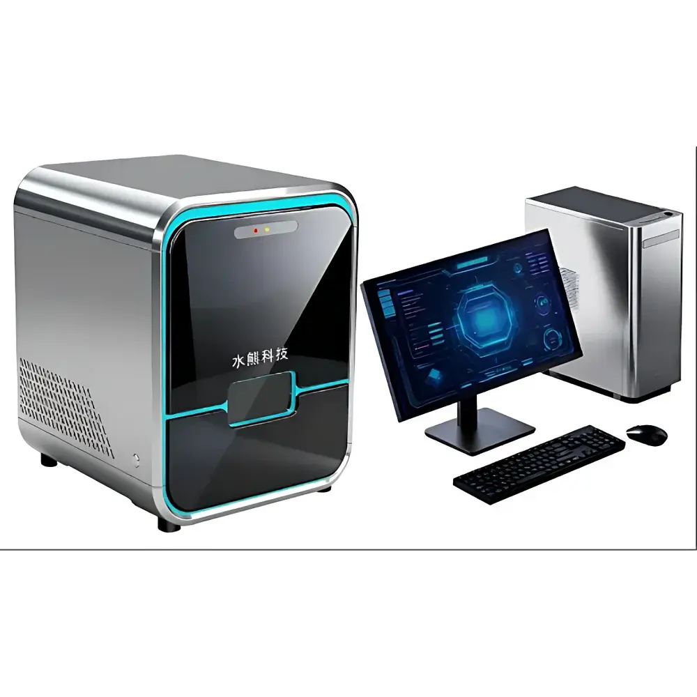
Water Bear Technology FC200 Laser Scanning Confocal Fluorescence Imager
| Brand | Water Bear Technology |
|---|---|
| Origin | Hebei, China |
| Manufacturer Type | Authorized Distributor |
| Country of Origin | China |
| Model | FC200 |
| Pricing | Available Upon Request |
| Imaging Principle | Confocal Laser Fluorescence Microscopy |
| Slide Format | Standard 25 × 75 mm Glass Slides |
| Maximum Scan Speed | 10 lines/sec |
| Optical Resolution | ≤5 µm (lateral, diffraction-limited) |
| Detection Mode | Multi-channel Fluorescence Intensity Quantification |
| Sample Platform | Integrated Microfluidic Chip Holder for Single-Cell Western Blot (scWB) Arrays |
Overview
The Water Bear Technology FC200 Laser Scanning Confocal Fluorescence Imager is a dedicated high-throughput, automated imaging platform engineered for quantitative single-cell proteomic analysis via confocal fluorescence detection. It operates on the principle of point-scanning confocal microscopy—using a focused laser beam and spatially synchronized pinhole detection to reject out-of-focus light, thereby enhancing axial resolution and signal-to-noise ratio (SNR) in thick or densely labeled samples. Unlike widefield systems, the FC200 achieves optical sectioning capability without physical sectioning, enabling precise quantification of fluorescently labeled proteins directly from intact microfluidic chips containing immobilized single cells. Designed specifically for single-cell Western blot (scWB) workflows, the system captures spatially registered fluorescence intensity data across multiplexed antibody panels, supporting both qualitative pattern recognition and absolute intensity-based relative quantification at the individual cell level.
Key Features
- Full-slide automated scanning compatible with standard 25 × 75 mm microscope slides, including custom microfluidic chip carriers pre-aligned for scWB arrays.
- High-speed galvanometric scanning engine delivering up to 10 lines per second—optimized for rapid acquisition of large-area, multi-channel fluorescence datasets without compromising lateral resolution.
- Confocal optical architecture with adjustable pinhole diameter and spectral separation filters, ensuring consistent depth discrimination and minimal crosstalk between fluorophores (e.g., FITC, TRITC, Cy5).
- Onboard fluorescence intensity quantification algorithm trained on validated scWB reference standards; supports background subtraction, pixel-wise normalization, and spot-level intensity integration.
- Integrated thermal and mechanical stabilization subsystems to maintain focus stability during extended acquisitions (>30 min), critical for longitudinal single-cell tracking experiments.
- Modular excitation source configuration supporting 405 nm, 488 nm, 561 nm, and 640 nm lasers—enabling four-color simultaneous or sequential detection aligned with common antibody conjugates.
Sample Compatibility & Compliance
The FC200 accommodates standardized glass substrates and proprietary microfluidic chips fabricated from cyclic olefin copolymer (COC) or fused silica—materials selected for low autofluorescence and compatibility with common fixation (paraformaldehyde), permeabilization (Triton X-100), and blocking reagents. The system supports formalin-fixed paraffin-embedded (FFPE) tissue sections when mounted on compatible adhesive slides, though optimal performance is achieved with fresh-frozen or chemically fixed single-cell suspensions processed via scWB protocols. All hardware and firmware comply with IEC 61000-6-3 (EMC emissions) and IEC 61000-6-2 (immunity) standards. Data handling adheres to ALCOA+ principles (Attributable, Legible, Contemporaneous, Original, Accurate, Complete, Consistent, Enduring, Available); audit trails and user access logs are configurable to support GLP and GMP environments where required.
Software & Data Management
The FC200 is operated via WB-ScanSuite v3.x—a Windows-based application developed in compliance with FDA 21 CFR Part 11 requirements for electronic records and signatures. Core modules include Acquisition Control, Multi-channel Registration Engine, Spot Detection & Intensity Mapping, and Batch Analysis Pipeline. Raw image data is stored in OME-TIFF format with embedded metadata (objective magnification, laser power, PMT gain, exposure time, channel assignment). Export options include CSV tables of normalized intensity values per detected cell, annotated TIFF overlays, and JSON-formatted metadata packages compatible with downstream analysis in Python (Scanpy, scikit-image) or R (Seurat, flowCore). Software validation documentation—including IQ/OQ/PQ test scripts—is available upon request for regulated laboratory deployment.
Applications
- Single-cell protein expression profiling in oncology research, including intra-tumoral heterogeneity mapping of kinase activation states (e.g., pERK, pAKT) and immune checkpoint markers (PD-L1, CTLA-4).
- Neuroscience applications involving primary neuron cultures or iPSC-derived neural progenitors, quantifying synaptic protein co-expression (e.g., PSD95/Synapsin) at subcellular resolution.
- Immunophenotyping of rare circulating tumor cells (CTCs) or antigen-specific T-cell subsets using multiplexed surface and intracellular marker panels.
- Clinical assay development for companion diagnostics requiring quantitative protein-level stratification—particularly where RNA-based methods lack functional correlation.
- Forensic toxicology and trace biological evidence analysis, leveraging ultra-low detection thresholds (<500 molecules/cell equivalent) enabled by high-SNR confocal detection.
FAQ
What sample preparation protocols are recommended for optimal FC200 performance?
Standard scWB workflows—including cell lysis on-chip, electrophoretic separation, antibody incubation, and fluorescent secondary detection—are validated. FFPE sections require antigen retrieval optimization prior to staining.
Does the FC200 support time-lapse imaging of live cells?
No—the system is optimized for fixed-sample, high-SNR confocal scanning. Live-cell imaging requires environmental chamber integration not included in base configuration.
Can third-party antibodies be used with the FC200’s quantification pipeline?
Yes—provided lot-specific titration curves and isotype controls are supplied to calibrate intensity response functions within WB-ScanSuite.
Is remote operation supported?
Yes—via secure RDP or VNC connections; however, raw data transfer must occur over local network due to file size constraints (>2 GB per full-slide scan).
How is calibration maintained across instrument lifetime?
Annual optical alignment verification and PMT gain calibration using NIST-traceable fluorescent microspheres (e.g., TetraSpeck) are recommended and documented in the service logbook.
Related Products


