Xunshu MCN-3L Automated Micronucleus Analysis System for Erythrocytes
| Brand | Xunshu |
|---|---|
| Origin | Zhejiang, China |
| Manufacturer Type | Original Equipment Manufacturer (OEM) |
| Country of Origin | China |
| Model | MCN-3L |
| Pricing | Upon Request |
Overview
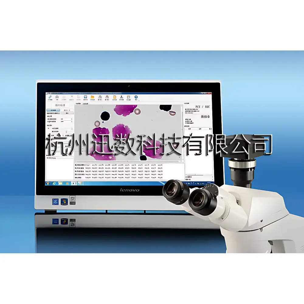
The Xunshu MCN-3L Automated Micronucleus Analysis System is a dedicated digital image analysis platform engineered for regulatory-compliant genotoxicity assessment in mammalian erythrocytes. It implements standardized in vivo micronucleus testing protocols per OECD Test Guideline 474, ISO 10993-3, and ICH S2(R2), focusing on the detection of micronuclei in polychromatic erythrocytes (PCEs) from rodent bone marrow or peripheral blood smears stained with Giemsa. The system operates on a dual-stage computational workflow: first, machine learning–driven classification of erythrocyte subtypes (PCE vs. normochromatic erythrocytes, NCE) using morphological and chromatic features; second, high-fidelity micronucleus identification based on strict geometric and optical criteria—including size (1/20 to 1/5 of PCE diameter), circularity, chromatin homogeneity with main nucleus, and edge smoothness. Its core imaging engine integrates real-time USB 3.0 video capture with a scientific-grade Sony ExView HAD CCD sensor (ICX694AQG(C), 6.0 MP, 1-inch format), enabling large-field-of-view acquisition at 2748 × 2200 resolution without stage scanning—reducing sampling bias and operator fatigue.
Key Features
- AI-Powered Cell Classification: Trained on representative datasets of Giemsa-stained PCEs and NCEs, the system performs unsupervised feature extraction and pattern recognition to achieve >98% classification accuracy under validated conditions.
- Resonance-Based Image Processing: Proprietary stochastic resonance algorithm enhances low-contrast micronucleus signals against heterogeneous cytoplasmic background, improving detection sensitivity without compromising specificity.
- High-Throughput Quantification: Processes ≥2000 PCEs per sample in ≤60 seconds; computes PCE/NCE ratio and micronucleus frequency (‰) automatically, with full traceability of each classified cell.
- Dual-Software Architecture: Integrates MCN-3L Micronucleus Analysis Module and MIC Microscopic Imaging & Counting Module—supporting clonal colony counting, multi-field particle statistics, and morphometric profiling.
- FDA 21 CFR Part 11–Ready Audit Trail: Immutable electronic logs record timestamped user actions—including login/logout, parameter changes, result edits, data exports, and account modifications—with role-based access control (Administrator/Operator tiers).
- Calibration-Traceable Metrology: On-system digital micrometer enables ISO/IEC 17025–aligned measurements of cell diameter, area, perimeter, circularity, and angular orientation; supports user-defined calibration standards.
Sample Compatibility & Compliance
The MCN-3L is validated for use with Giemsa-stained cytospin or smear preparations from mouse/rat bone marrow aspirates and peripheral blood. It complies with GLP principles for nonclinical laboratory studies (OECD Series on Principles of Good Laboratory Practice) and supports audit readiness for regulatory submissions to FDA, EMA, PMDA, and NMPA. All analytical outputs—including raw image metadata, classification confidence scores, and statistical summaries—are stored in encrypted local databases with SHA-256 hashing. PDF reports generated via the system embed digital signatures and are functionally identical to underlying electronic records—ensuring ALCOA+ (Attributable, Legible, Contemporaneous, Original, Accurate, Complete, Consistent, Enduring, Available) data integrity.
Software & Data Management
The MCN-3L runs on Windows 11 Pro (pre-installed on Lenovo all-in-one workstation: Intel Core i5, 16 GB RAM, 1 TB HDD + 256 GB SSD, 27″ FHD display). Both MCN and MIC modules support DICOM-compliant image import/export and native TIFF/PNG/JPEG handling. Image databases accommodate up to five experimental groups (high/medium/low dose, positive/negative control), 50 slides, and 10,000 fields of view—with spatial indexing enabling pixel-accurate relocalization of any analyzed cell within its original acquisition frame. All quantitative results export to Excel (.xlsx) or PDF with embedded metadata (acquisition date, operator ID, microscope objective, magnification, exposure time). Electronic records are write-protected post-analysis; deletion requires dual-administrator approval and generates an auditable log entry.
Applications
- Regulatory genotoxicity screening of pharmaceutical candidates, agrochemicals, and industrial chemicals per OECD TG 474.
- Internal safety pharmacology studies supporting IND/IMPD dossiers.
- Quality control of biologics and cell therapies where chromosomal instability must be monitored.
- Academic research in radiation biology, oxidative stress response, and DNA repair mechanisms.
- Automated colony-forming unit (CFU) enumeration in hematopoietic progenitor assays.
- Morphometric analysis of micronucleated cells in environmental toxicology (e.g., heavy metal exposure models).
FAQ
Does the MCN-3L require integration with a specific microscope model?
No—the system interfaces with any upright or inverted microscope equipped with a trinocular port and C-mount adapter; no proprietary hardware coupling is needed.
Can the software distinguish micronuclei from apoptotic bodies or staining artifacts?
Yes—through combined constraints on size, shape factor (circularity ≥0.75), chromatin density uniformity, and spatial isolation from nuclear membrane remnants.
Is remote access or network deployment supported?
Local area network (LAN) deployment is supported for centralized database management; remote desktop access must comply with institutional IT security policies and is not enabled by default.
What validation documentation is provided?
Xunshu supplies IQ/OQ documentation templates, system suitability test protocols, and a comprehensive User Requirement Specification (URS) aligned with ISO/IEC 17025 and ASTM E2500.
How frequently must the AI models be retrained?
Model drift is mitigated through periodic recalibration using reference slide sets; annual retraining is recommended when introducing new staining protocols or species-specific samples.


