
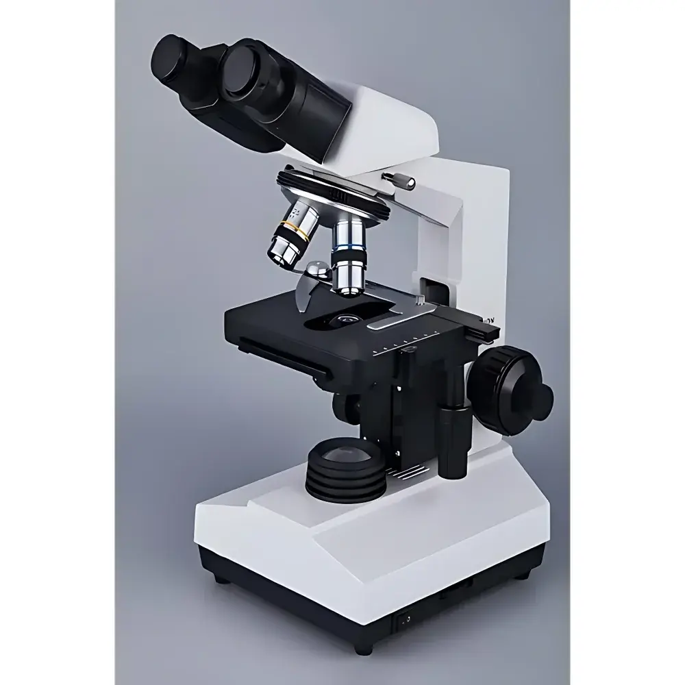

Yoke XSP-8CA Dual-Eyepiece Biological Microscope
| Brand | Yoke |
|---|---|
| Origin | Shanghai, China |
| Model | XSP-8CA |
| Microscope Type | Digital Biological Microscope |
| Eyepiece Configuration | Binocular |
| Objective Lens System | Finite-conjugate Achromatic Objectives (Standard: 4×, 10×, 40× spring-loaded |
| Optional | 100× oil-immersion spring-loaded) |
| Eyepieces | Wide-field WF10× and WF16× (pair included) |
| Head | Interchangeable铰链式 binocular head with 30° inclined eyepieces |
| Nosepiece | Quadruple revolving nosepiece |
| Stage | Double-layer mechanical stage (135 × 140 mm) with coaxial X–Y controls |
| Focus Mechanism | Coaxial coarse/fine focusing |
| fine focus graduation | 0.002 mm |
| Condenser | Abbe condenser, NA = 1.25, with iris diaphragm |
| Illumination | High-brightness, continuously adjustable LED (12 V DC, powered via wide-input 100–230 V AC adapter) |
| Compliance | Designed for routine laboratory use in teaching, clinical screening, and life science research environments |
| Software Compatibility | Compatible with standard USB digital imaging modules (not included) |
Overview
The Yoke XSP-8CA Dual-Eyepiece Biological Microscope is a robust, entry-to-mid-tier upright microscope engineered for routine brightfield observation in academic teaching laboratories, clinical cytology workflows, and applied life science research settings. It employs a finite-conjugate optical pathway with achromatic objective lenses—designed to correct for chromatic aberration at two wavelengths and spherical aberration at the central wavelength—ensuring high-fidelity monochrome image fidelity across the visible spectrum (400–700 nm). The instrument’s mechanical architecture follows ergonomic design principles: the 30° inclined binocular head reduces cervical strain during extended observation sessions, while the coaxial coarse/fine focusing system—with calibrated fine-focus graduations of 0.002 mm—enables precise Z-axis positioning required for serial section analysis or multiplane documentation. Its wide-input power supply (100–230 V AC) ensures stable 12 V DC operation of the high-luminance LED illuminator, eliminating filament degradation and thermal drift associated with halogen sources.
Key Features
- Finite-conjugate achromatic objective lens set (standard: 4×, 10×, 40× spring-loaded; optional oil-immersion 100× objective compliant with ISO 8578:2017 for immersion medium refractive index matching)
- Wide-field eyepieces (WF10× and WF16×, supplied as matched pair) providing field numbers of 18 mm and 16 mm respectively, supporting extended visual scanning without frequent repositioning
- Quadruple revolving nosepiece with precision ball-bearing rotation and positive click-stop indexing for repeatable objective alignment
- Double-layer mechanical stage (135 × 140 mm) with low-backlash lead-screw drives and vernier scales for sub-millimeter specimen navigation
- Abbe condenser (NA = 1.25) equipped with an iris diaphragm and centering screws—enabling Köhler illumination setup per ISO 10934-1:2001 standards
- LED illumination system featuring continuous intensity control (0–100%), flicker-free output, and >50,000-hour rated lifetime—reducing maintenance frequency and energy consumption relative to tungsten-halogen systems
Sample Compatibility & Compliance
The XSP-8CA accommodates standard 1″ × 3″ glass slides (76 × 26 mm) and 24 × 50 mm coverslips. Its stage aperture and condenser height allow unobstructed observation of stained histological sections (e.g., H&E, Gram), live unstained protozoa in hanging-drop preparations, and fixed bacterial smears. While not certified for ISO/IEC 17025 accreditation as a metrological instrument, its optical performance aligns with ASTM E287-21 (Standard Specification for Microscopes Used in Clinical Microscopy) for resolution validation using USAF 1951 test targets. The microscope supports GLP-compliant documentation when integrated with FDA 21 CFR Part 11–enabled digital imaging software (e.g., via third-party USB cameras with audit-trail logging).
Software & Data Management
The XSP-8CA is hardware-compatible with industry-standard USB 2.0/3.0 digital microscopy cameras (e.g., 5 MP CMOS sensors). When paired with validated acquisition software—such as Olympus cellSens, Leica LAS X Core, or open-source alternatives like MicroManager—the system supports time-lapse capture, multi-channel overlay (with filter cube adapters), and metadata embedding (objective magnification, exposure time, illumination intensity). All digital outputs retain EXIF-compliant headers, facilitating traceability in teaching labs and quality-controlled environments where image provenance is required per ISO/IEC 17025 clause 7.5.2.
Applications
- Undergraduate and graduate biology instruction: mitosis staging in onion root tip squashes, blood smear differential counts, fungal hyphae morphology
- Clinical pre-screening: urinalysis sediment examination, wet-mount evaluation for vaginal flora, rapid malaria parasite detection in Giemsa-stained thin films
- Agricultural diagnostics: nematode identification in soil extracts, pollen grain viability assessment, seed coat microstructure analysis
- Industrial QC: inspection of textile fiber integrity, pharmaceutical tablet coating uniformity (via surface reflection mode with optional darkfield stop), and printed circuit board solder joint verification
FAQ
Is the XSP-8CA compatible with oil immersion objectives?
Yes—the standard configuration includes a spring-loaded 100× oil-immersion objective (NA 1.25), and the Abbe condenser supports full numerical aperture utilization when used with Type A immersion oil (nD = 1.515 ± 0.001).
Can this microscope be used for photomicrography?
Yes—its trinocular port option (available as accessory) enables simultaneous visual observation and camera coupling; USB-digital cameras mount directly to the C-mount adapter (0.5× relay lens recommended for optimal field coverage).
Does the LED illumination support Köhler alignment?
Yes—the condenser includes centering screws and a field diaphragm, permitting full Köhler illumination setup per ISO 10934-1:2001 to optimize contrast and resolution.
What is the maximum useful magnification achievable with this system?
Under ideal conditions (100× objective + 10× eyepiece), total magnification reaches 1000×; however, practical resolution is limited by the objective’s NA (1.25) and visible light wavelength, yielding ~0.2 µm theoretical resolution per Rayleigh criterion.
Is service and calibration support available outside mainland China?
Yoke provides OEM technical documentation and firmware updates globally; third-party service partners in EU and North America offer preventive maintenance and optical recalibration per ISO 9001-certified procedures.


