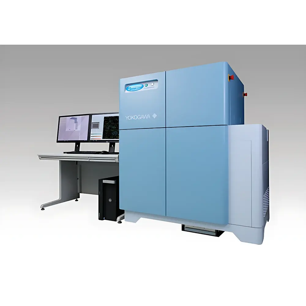
Yokogawa CellVoyager CV8000 High-Content Screening System
| Brand | Yokogawa |
|---|---|
| Origin | Japan |
| Manufacturer | Yokogawa Electric Corporation |
| Type | CellVoyager CV8000 |
| Excitation Wavelengths | 405 nm, 445 nm, 488 nm, 561 nm, 640 nm (solid-state lasers, up to 5 channels) |
| Objective Lenses | 2×–60× dry, phase contrast, water-immersion, and long-working-distance options |
| Camera | Up to four high-sensitivity sCMOS cameras (2000 × 2000 pixels, 6.5 µm pixel size) |
| Autofocus | Dual-mode (laser-based and image-based) |
| Confocal Unit | Dual Nipkow disk scanning system with micro-lens enhancement |
| pinhole disk options | 25 µm or 50 µm, auto-exchangeable |
| Sample Formats | 6–1536-well plates, slides |
| Environmental Control | On-stage incubator with temperature (35–40 °C), CO₂ (5%), and humidity regulation |
| Robotic Liquid Handler | Optional 96-/384-tip disposable pipetting module with barcode reader support |
| Software | CellPathfinder (AI-powered analysis, label-free CE brightfield reconstruction, 3D segmentation, machine learning & deep learning modules) |
| Data Output | 16-bit TIFF/PNG images |
| Compliance | Designed for GLP/GMP-aligned workflows |
Overview
The Yokogawa CellVoyager CV8000 is a fully integrated, high-content screening (HCS) platform engineered for quantitative, longitudinal, and physiologically relevant phenotypic analysis of living cells and 3D tissue models. At its core lies Yokogawa’s proprietary dual-disk spinning confocal scanning technology—combining a microlens-enhanced Nipkow disk with a precision-aligned pinhole array disk—to deliver optical sectioning at speeds exceeding 1,000 frames per second while minimizing phototoxicity and photobleaching. Unlike conventional widefield or point-scanning systems, the CV8000 achieves true confocality across large fields of view and thick specimens (e.g., organoids, spheroids, and co-cultures) without compromising temporal resolution or signal fidelity. Its on-stage environmental chamber maintains stable CO₂ (5%), temperature (35–40 °C), and humidity levels for uninterrupted live-cell imaging over periods exceeding 72 hours—validated against standard incubators for uniform proliferation kinetics across 96-well plates (excluding corner wells). This architecture bridges the “valley of death” in drug discovery by enabling concurrent high-throughput screening and high-fidelity phenotypic profiling under near-physiological conditions.
Key Features
- Dual-Nipkow Confocal Scanning: Patented dual-disk configuration with interchangeable 25 µm and 50 µm pinhole arrays enables adaptive optical sectioning—optimized for either enhanced axial resolution (smaller pinholes) or improved signal-to-noise ratio (larger pinholes) depending on sample thickness and fluorophore brightness.
- Multi-Camera sCMOS Imaging: Up to four synchronized sCMOS cameras (2000 × 2000 pixels, 6.5 µm pixel pitch) allow simultaneous acquisition of four fluorescence channels plus brightfield or phase contrast—achieving full 96-well plate imaging in under 60 seconds at 4-color multiplexing.
- Water-Immersion Optics: Integrated 40× and 60× water-immersion objectives with active spherical aberration correction enable high-resolution imaging of cells in aqueous environments—critical for long-term observation of delicate primary cultures and 3D organoid models.
- On-Stage Incubation & Robotic Integration: Sealed, humidity-controlled stage incubator eliminates thermal drift and condensation. Optional integrated robotic liquid handler supports automated reagent addition (e.g., compound titration, stimulus application) during time-lapse acquisition—with precise timing, multi-step dispensing, and tip-tracking via barcode readers.
- Label-Free Phenotypic Profiling: CellPathfinder software reconstructs contrast-enhanced (CE) brightfield Z-stacks using computational deconvolution and AI-driven segmentation—enabling morphometric analysis of unlabeled iPSCs, neurons, or tumor spheroids without fixation, staining, or genetic modification.
Sample Compatibility & Compliance
The CV8000 accommodates diverse biological formats including 6–1536-well microplates, glass slides, and custom chambers. It is routinely deployed for adherent and suspension cultures, primary human cells, induced pluripotent stem cell (iPSC)-derived lineages, and complex 3D models such as cerebral organoids, tumor spheroids, and endothelial tubules. The system meets engineering and operational prerequisites for regulated environments: hardware design supports 21 CFR Part 11-compliant data governance (electronic signatures, audit trails, role-based access), and software validation packages are available for GLP and GMP workflows. All environmental parameters—including CO₂ concentration, temperature setpoints, and humidity—are logged continuously with timestamped metadata embedded in image headers and raw data files.
Software & Data Management
CellPathfinder serves as the unified analytical engine for the CV8000—providing modular, protocol-driven analysis from acquisition through visualization. Its workflow interface features drag-and-drop protocol assembly, template libraries for common assays (e.g., nuclear translocation, neurite outgrowth, mitotic index), and version-controlled script export. CE brightfield reconstruction leverages multi-Z plane acquisition to generate synthetic contrast images suitable for machine learning training. The optional Deep Learning Module integrates convolutional neural networks (CNNs) trained on user-annotated datasets to classify subtle morphological phenotypes—such as early apoptosis or cytoskeletal remodeling—with reproducibility exceeding manual scoring. Quantitative outputs (e.g., object count, texture metrics, spatial distribution statistics) are exported in CSV and HDF5 formats, linked directly to source images for traceable validation. CellLibrarian provides centralized metadata tagging, hierarchical project organization, and REST API access for integration with LIMS and ELN systems.
Applications
The CV8000 addresses critical bottlenecks in translational research and preclinical development. In neurodegenerative disease modeling, it quantifies dynamic changes in dendritic spine density and mitochondrial motility in iPSC-derived neurons over 7-day time courses. In immuno-oncology, it tracks T-cell infiltration, tumor cell killing kinetics, and PD-L1 membrane translocation in 3D co-culture spheroids. For toxicology screening, it performs multiparametric assessment of cardiomyocyte beating frequency, calcium transient amplitude, and sarcomere structure in hiPSC-derived cardiac microtissues. Its compatibility with FRET biosensors and Cell Painting protocols further extends utility into pathway-specific signaling dynamics and unbiased phenotypic fingerprinting. Academic labs leverage its open architecture for method development in CRISPR screening validation, while biopharma users deploy it in lead optimization campaigns requiring >10,000 compound evaluations with subcellular resolution.
FAQ
Does the CV8000 support true 3D volumetric imaging of thick specimens?
Yes—the dual-disk confocal architecture combined with motorized Z-focus and water-immersion optics enables high-fidelity optical sectioning of samples up to 200 µm thick, with demonstrated performance in organoid and tissue slice imaging.
Can CellPathfinder analyze unlabeled cells without fluorescent dyes or transfection?
Yes—its CE brightfield reconstruction algorithm converts multi-Z brightfield stacks into high-contrast synthetic images suitable for AI-based segmentation and feature extraction, eliminating labeling artifacts and variability.
Is the system compatible with existing laboratory automation infrastructure?
Yes—the CV8000 supports standard communication protocols (SLIM, RS-232, TCP/IP) and integrates with third-party robotic arms, plate stackers, and LIMS via Yokogawa’s OpenAPI framework.
What level of regulatory compliance does the software provide?
CellPathfinder supports 21 CFR Part 11 compliance through electronic signatures, immutable audit logs, and configurable user roles—validated documentation packages are available for IQ/OQ/PQ execution.
How is phototoxicity managed during extended live-cell experiments?
By combining low-light sCMOS detection, high-efficiency microlens-enhanced excitation, and adaptive pinhole selection, the CV8000 reduces effective laser dose by up to 70% compared to single-point scanning systems—enabling 72+ hour imaging of sensitive stem cell populations.


