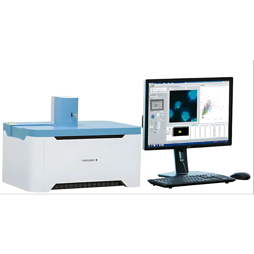
Yokogawa CQ1 Benchtop High-Content Analysis System
| Brand | Yokogawa |
|---|---|
| Origin | Japan |
| Model | CQ1 |
| Configuration | Benchtop, Motorized Stage with Environmental Chamber Option |
| Imaging Technology | Microlens-Enhanced Nipkow Disk Confocal |
| Max Acquisition Speed | 20 fps (optional) |
| Data Export Formats | FCS, CSV, ICE |
| Sample Compatibility | Standard Petri Dishes, Multiwell Plates (6–384-well), Tissue Slices, Spheroids, Colonies |
| Field of View | Wide-field tiling enabled |
| Light Source | Solid-state lasers (405/488/561/640 nm typical) |
| Detection | Back-illuminated EM-CCD or sCMOS camera (model-dependent) |
| Software Platform | CV8000-compatible analysis suite with open API |
Overview
The Yokogawa CQ1 Benchtop High-Content Analysis System is an engineered solution for quantitative, label-based 3D cellular phenotyping in adherent and semi-adherent biological specimens. It employs microlens-enhanced Nipkow disk confocal microscopy—a parallelized optical sectioning technique that combines high-speed raster scanning with inherently low phototoxicity and photobleaching. Unlike conventional flow cytometry, the CQ1 acquires spatially resolved, multi-parametric image data directly from intact culture vessels without enzymatic dissociation or suspension handling. This preserves native cell morphology, intercellular architecture, and microenvironmental context—critical for modeling tissue-level responses in spheroids, organoids, and histological sections. The system integrates a precision motorized stage with optional temperature, CO₂, and humidity control, enabling long-term live-cell imaging under physiologically relevant conditions. Its compact benchtop footprint eliminates requirements for dedicated darkrooms or vibration-isolated tables, making it deployable in standard biosafety cabinets and core imaging facilities alike.
Key Features
- Microlens-enhanced dual-disk scanning: A proprietary Nipkow disk containing ~20,000 pinholes is mechanically coupled with a matching microlens disk; each lens focuses excitation laser light onto its corresponding pinhole, enabling simultaneous multi-beam illumination and efficient light collection.
- Low-phototoxicity acquisition: Multi-beam illumination distributes excitation energy across multiple focal points, permitting high signal-to-noise ratio imaging at sub-mW laser power levels—essential for extended time-lapse studies of sensitive primary cells and stem cell derivatives.
- Wide-field tiling & large-sample support: Automated stage navigation enables seamless stitching of high-resolution z-stacks across large-area samples including 10 cm Petri dishes, whole-tissue sections, and multi-spheroid arrays.
- Real-time feature extraction: On-the-fly quantification of morphological, intensity-, texture-, and relational parameters (e.g., nuclear-cytoplasmic ratio, mitochondrial network fragmentation, cell-cell contact area) with immediate visualization via interactive scatter plots and histograms.
- Open data architecture: Native export to FCS (Flow Cytometry Standard), CSV, and ICE (Image Cytometry Exchange) formats ensures compatibility with third-party analysis platforms such as CellProfiler, Ilastik, Python-based scikit-image pipelines, and commercial HCA software suites.
Sample Compatibility & Compliance
The CQ1 accommodates standard cell culture formats—including 6–384-well plates, chambered coverslips, glass-bottom dishes, and custom-inserted tissue slices—without modification. Its non-invasive imaging paradigm aligns with Good Laboratory Practice (GLP) and Good Manufacturing Practice (GMP) principles by eliminating sample manipulation artifacts associated with trypsinization or centrifugation. While not certified as a medical device, the system supports experimental workflows compliant with ISO 20387 (biobanking), ASTM E2925 (standard guide for high-content screening assay development), and USP <1043> (cellular assays). Audit trail functionality, user access controls, and electronic signature readiness are available via optional CV8000-derived software modules meeting FDA 21 CFR Part 11 requirements.
Software & Data Management
The CQ1 operates with Yokogawa’s unified HCA software platform—derived from the CV8000 architecture—featuring modular acquisition, segmentation, and analysis modules. Image processing leverages adaptive thresholding, machine learning–assisted object classification (e.g., distinguishing apoptotic vs. necrotic nuclei), and batch-mode pipeline scripting. All raw and processed datasets retain full metadata (acquisition time, objective magnification, z-step size, laser power, gain settings), ensuring traceability from statistical output back to individual pixel values. Data storage follows FAIR principles (Findable, Accessible, Interoperable, Reusable), with DICOM-SR export capability for integration into institutional LIMS or ELN systems.
Applications
- 3D tumor spheroid drug response profiling: Quantifying viability gradients, hypoxia marker distribution, and invasion front dynamics over 72+ hour time courses.
- Stem cell differentiation monitoring: Tracking lineage-specific marker expression, colony morphology evolution, and mitotic index changes without endpoint fixation.
- Neuroscience co-culture analysis: Measuring synaptic puncta density, neurite outgrowth complexity, and glial activation states in mixed cortical cultures.
- Microbial colony phenotyping: Assessing antibiotic-induced morphological heterogeneity and persister cell subpopulations within biofilm-like structures.
- Toxicology screening: Detecting subtle cytoskeletal disruption, lysosomal accumulation, or mitochondrial membrane potential loss prior to overt cytotoxicity.
FAQ
Does the CQ1 require specialized training to operate?
Basic operation—plate loading, protocol selection, and data export—can be performed by trained laboratory technicians after a two-day on-site workshop. Advanced analysis pipeline development benefits from familiarity with image analysis concepts but does not require programming expertise due to the GUI-driven workflow builder.
Can the CQ1 be integrated into automated liquid handling or robotic systems?
Yes. The system features industry-standard RS-232, Ethernet, and TTL I/O interfaces, and supports communication via Yokogawa’s OpenAPI (RESTful HTTP endpoints), enabling bidirectional handshake with deck management software from Hamilton, Tecan, or Beckman Coulter platforms.
Is z-stack reconstruction compatible with deconvolution algorithms?
Raw TIFF stacks include precise z-position metadata and PSF calibration files; these are fully compatible with iterative deconvolution tools such as Huygens Professional, AutoQuant X, or Python-based DeconvolutionLab2.
What maintenance is required for the Nipkow disk assembly?
The dual-disk mechanism is sealed and factory-aligned; no user-serviceable parts exist. Annual preventive maintenance includes laser power calibration, stage positional accuracy verification, and camera quantum efficiency validation—performed by Yokogawa-certified field service engineers.
How does the CQ1 compare to point-scanning confocal systems in resolution and speed?
Lateral resolution (~250 nm) matches diffraction-limited point-scanners using the same objectives; axial resolution (~600 nm) is comparable at equivalent NA. However, the CQ1 achieves up to 20× faster volumetric acquisition than resonant-galvo or AOTF-based scanners—particularly advantageous for kinetic assays requiring >100 time points per condition.


