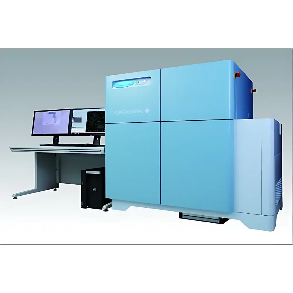
Yokogawa CV8000 High-Content Screening System
| Brand | Yokogawa |
|---|---|
| Origin | Japan |
| Model | CV8000 |
| Type | Automated Confocal High-Content Imaging Platform |
| Application Domain | Phenotypic Screening, 3D Cell Culture Analysis, Live-Cell Kinetic Imaging |
| Regulatory Context | Designed for GLP-compliant workflows |
Overview
The Yokogawa CV8000 High-Content Screening (HCS) System is an automated, confocal-based imaging platform engineered for quantitative, multi-parametric phenotypic analysis in drug discovery and functional cell biology. It operates on the principle of spinning-disk confocal microscopy, enabling optical sectioning with high signal-to-noise ratio while minimizing phototoxicity—critical for longitudinal live-cell assays. Unlike widefield HCS systems, the CV8000 integrates Yokogawa’s proprietary CSU-W1 spinning-disk unit with water-immersion objectives (e.g., 20×/1.0 NA, 40×/1.15 NA), delivering subcellular resolution (lateral resolution ≤ 280 nm at 488 nm excitation) and consistent Z-stack acquisition across large fields of view. Its architecture supports seamless integration into automated screening pipelines, including compatibility with standard microplate formats (96-, 384-, and 1536-well), robotic deck interfaces, and environmental control modules (37 °C, 5% CO₂, humidity regulation). The system is purpose-built to bridge the “valley of death” in preclinical development by enabling physiologically relevant assay models—such as spheroids, organoids, and co-cultures—without sacrificing throughput.
Key Features
- High-speed confocal imaging: Dual-camera configuration (up to four synchronized sCMOS cameras optional) enables parallel multi-channel acquisition with frame rates up to 30 fps per channel at full resolution (2048 × 2048 pixels).
- Water-immersion objective optics: Reduces spherical aberration in thick specimens; maintains focus stability during extended time-lapse imaging of 3D cultures.
- Integrated environmental control: Motorized stage with on-stage incubation chamber ensures precise thermal and gaseous regulation (±0.2 °C temperature stability, CO₂ feedback control) throughout acquisition.
- Automated liquid handling: On-deck pipetting module supports reagent addition, medium exchange, and compound dilution directly within the imaging workflow—reducing manual intervention and cross-plate variability.
- Modular optical path: Configurable laser lines (405, 488, 561, 640 nm) and emission filters support multiplexed detection of ≥6 fluorescent labels per field, including FRET and spectral unmixing-ready setups.
- Robust mechanical design: Vibration-damped optical table integration, active focus stabilization (Z-drift compensation < 50 nm/hr), and ISO Class 5 cleanroom-compatible enclosure options.
Sample Compatibility & Compliance
The CV8000 accommodates diverse biological samples: adherent and suspension cells, primary isolates, stem-cell-derived organoids, hydrogel-embedded spheroids (up to 500 µm diameter), and tissue explants cultured in specialized inserts. Its non-invasive imaging modality avoids fixation artifacts, supporting kinetic assays spanning hours to days. From a regulatory standpoint, the system meets essential requirements for pharmaceutical R&D environments: hardware design aligns with IEC 61000-6-2/6-4 electromagnetic compatibility standards; firmware supports user access levels, electronic signatures, and audit trail generation per FDA 21 CFR Part 11 when paired with validated CellPathfinder v5.0+ software. It is routinely deployed in laboratories operating under GLP and early-phase GMP-aligned quality systems, with documentation packages available for IQ/OQ/PQ execution.
Software & Data Management
CellPathfinder software serves as the native analysis engine, offering both rule-based segmentation and AI-driven object recognition. Its deep learning module (trained on >2 million manually annotated cellular structures) enables robust identification of morphologically heterogeneous targets—e.g., neurite outgrowth in iPSC-derived neurons, mitochondrial network fragmentation, or nuclear translocation events—without requiring user-defined thresholds. Batch processing supports parallel analysis of >100 plates per run, with metadata tagging compliant with MIAME and MIAPE guidelines. Raw data (OME-TIFF format) and processed results (CSV, HDF5) are exportable to LIMS or ELN platforms via RESTful API. Version-controlled analysis pipelines ensure reproducibility across sites and operators, and software validation kits—including test scripts, traceability matrices, and UAT protocols—are provided for regulated environments.
Applications
- Phenotypic screening of compound libraries against disease-relevant cellular models (e.g., tau aggregation in Alzheimer’s iPSC-neurons, lipid droplet accumulation in NAFLD hepatocytes).
- Functional cytotoxicity assessment using multiplexed readouts: caspase-3 activation + mitochondrial membrane potential + nuclear morphology.
- 3D tumor spheroid invasion assays with collagen-I matrix embedding and time-resolved tracking of collective cell migration.
- Stem cell differentiation monitoring via label-free phase contrast + fluorescent lineage reporters over 14-day timelines.
- Host-pathogen interaction studies: intracellular bacterial load quantification in macrophages coupled with cytokine secretion profiling via secreted antibody capture arrays.
FAQ
Does the CV8000 support real-time autofocus during long-term time-lapse imaging?
Yes—the system employs a near-infrared reflection-based focus lock mechanism that continuously monitors coverslip position with nanometer-level sensitivity, compensating for thermal drift and medium evaporation without perturbing visible-light imaging channels.
Can CellPathfinder be validated for use in GxP-regulated environments?
Yes—Yokogawa provides a comprehensive Computer System Validation (CSV) support package, including risk assessments, requirement specifications, test protocols, and summary reports aligned with Annex 11 and ALCOA+ principles.
Is the CV8000 compatible with third-party microfluidic chips or custom culture substrates?
The stage accommodates standard footprint adapters (e.g., for ibidi µ-Slides or Mimetas OrganoPlates); custom mechanical interfaces can be developed through Yokogawa’s Engineering Services group upon feasibility review.
What is the maximum Z-stack depth achievable in a single acquisition cycle?
Up to 100 optical sections at 0.5 µm intervals can be acquired per field in < 90 seconds using the 40× water objective, with automatic intensity normalization across slices to maintain quantitative fidelity.
How does the CV8000 handle photobleaching during multi-day experiments?
It implements adaptive illumination control: laser power and exposure time are dynamically adjusted per channel and Z-plane based on real-time histogram feedback, preserving fluorophore integrity while maintaining SNR above 20 dB across 72-hour acquisitions.
Related Products


