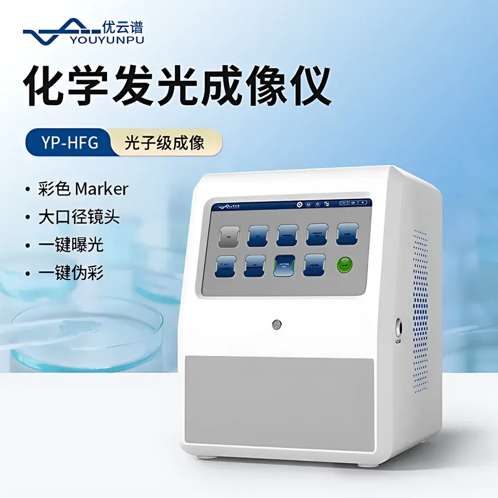
Youyunpu YP-HFG Chemiluminescence and Fluorescence Imaging System
| Brand | Youyunpu |
|---|---|
| Origin | Shandong, China |
| Manufacturer Type | Direct Manufacturer |
| Product Origin | Domestic (China) |
| Model | YP-HFG |
| Price | USD 12,300 (approx. ¥88,000) |
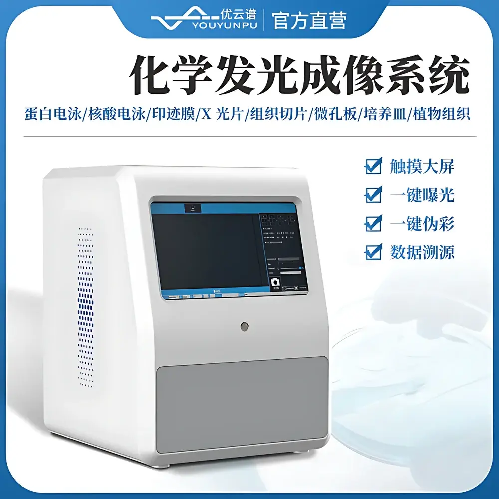
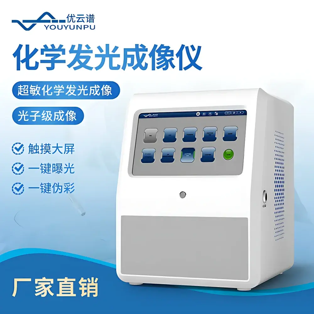
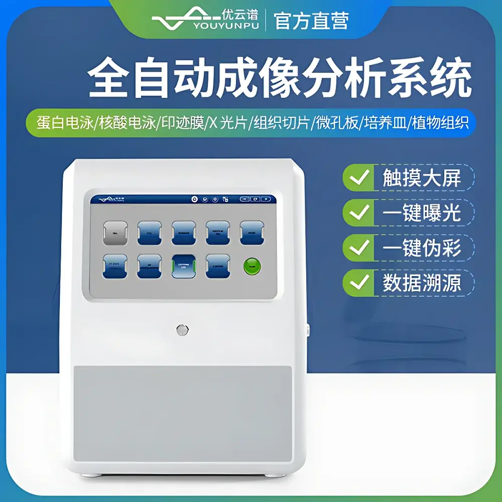
| Touchscreen | 13.3-inch capacitive |
| Memory | 16 GB RAM |
| Storage | 256 GB SSD |
| Camera Sensor | Back-illuminated scientific CMOS, 1-inch format |
| Dynamic Range | ≥4.8 decades |
| Bit Depth | 16-bit |
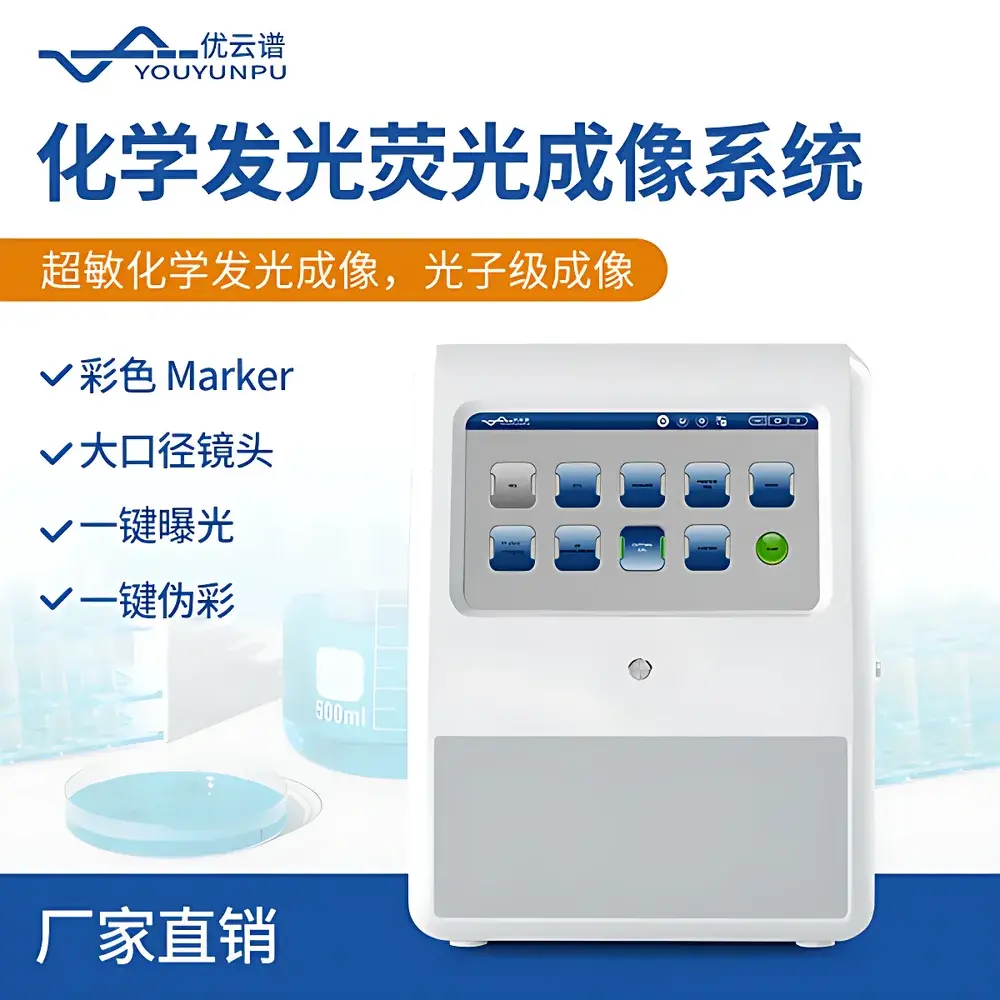
| Lens | Macro f/0.95 (optional f/0.8) |
| Cooling | −45°C below ambient, stabilization in ≤3 min |
| Binning Modes | 1×1 to 16×16 |
| Max Sample Area | >20 × 18 cm |
| Filter Wheel | 8-position, standard 595 nm (≥90% transmission) |
| Excitation Sources | Reflected white light, transmitted UV (365 nm standard |
Overview
The Youyunpu YP-HFG Chemiluminescence and Fluorescence Imaging System is a fully integrated, automated optical imaging platform engineered for high-sensitivity detection of chemiluminescent, fluorescent, and visible-light signals across diverse biological specimens. Built upon photon-counting imaging architecture, the system combines a back-illuminated 1-inch scientific CMOS sensor with ultra-fast f/0.95 macro optics and active thermoelectric cooling (−45°C below ambient), enabling quantitative capture of weak ECL signals—such as those from low-abundance phosphorylated ERK in Western blots—with detection limits down to 5.2 pg per band. Its ≥4.8-decade dynamic range ensures simultaneous linear quantification of both saturated and sub-picomolar signal intensities within a single exposure, eliminating the need for multiple exposures or manual band re-imaging. Designed for compliance with GLP/GMP-aligned laboratory workflows, the YP-HFG supports traceable, auditable image acquisition and analysis without reliance on proprietary cloud infrastructure.
Key Features
- Photon-level imaging engine with real-time noise suppression, delivering 2–3× faster acquisition (40–60 s vs. conventional 2-min exposures) while preserving quantitative linearity.
- 1-inch back-illuminated CMOS detector with selectable binning (1×1 to 16×16), enabling adaptive sensitivity tuning for ultra-weak signal detection or high-resolution spatial mapping.
- f/0.95 (or optional f/0.8) large-aperture macro lens providing >2.5× higher photon throughput than standard f/1.4 lenses—critical for preserving signal-to-noise ratio in low-light ECL applications.
- Automated motorized focus with <2 s convergence time, eliminating manual focusing variability and ensuring repeatable focal plane alignment across multi-sample runs.
- Intelligent auto-exposure algorithm that dynamically selects optimal integration time based on real-time preview histogram analysis—no user-defined thresholding required.
- Integrated color marker synthesis: automatic overlay of calibrated RGB ladder images onto monochrome chemiluminescent data, supporting intuitive molecular weight estimation without post-acquisition software stitching.
- AI-assisted batch naming: single-label propagation across replicate or time-series acquisitions using contextual metadata (e.g., sample ID, blot number, antibody target), reducing clerical error risk.
- Full audit trail embedding: each TIFF or PNG output file contains embedded EXIF metadata including acquisition timestamp, exposure parameters (gain, integration time, binning), user ID, and instrument firmware version.
- One-click pseudo-color mapping with 12 preset LUTs (e.g., Fire, Jet, Hot, Rainbow), enabling immediate visual discrimination of relative signal intensity gradients across membranes or gels.
- Onboard image processing suite with non-destructive editing: region-of-interest cropping, 90° rotation, overexposure pixel masking, gamma correction, and background subtraction using rolling-ball or polynomial fitting algorithms.
Sample Compatibility & Compliance
The YP-HFG accommodates standard and custom-format specimens—including SDS-PAGE gels, agarose/nucleic acid gels, nitrocellulose/PVDF transfer membranes, X-ray film, histological tissue sections (frozen or FFPE), microtiter plates (6–384-well), Petri dishes, and whole-plant organs (e.g., Arabidopsis leaves for bioluminescence monitoring). Its >20 × 18 cm imaging area supports full-gel visualization without tiling. All excitation sources comply with IEC 62471 photobiological safety standards. UV transillumination modules meet ISO 15223-1 labeling requirements for Class 3B laser product equivalents. Image metadata structures conform to NIH-approved MIAME-compliant extensions, facilitating submission to public repositories such as PRIDE or GEO. The system’s deterministic exposure control and hardware-based gain calibration support validation under FDA 21 CFR Part 11 when paired with institutional electronic signature policies.
Software & Data Management
The embedded Linux-based operating system hosts two tightly coupled applications: ImageCapture Pro (acquisition) and QuantStudio Analyst (analysis). ImageCapture Pro features touchscreen-optimized UI with gesture navigation, real-time histogram overlays, and overexposure warning overlays (highlighting clipped pixels in red). It supports five exposure modes: Auto, Manual, Accumulated (frame-averaged), Total Accumulated (summed), and User-Defined Multi-Step protocols. QuantStudio Analyst performs lane/band detection via adaptive edge detection, background modeling, and volume-integrated densitometry. Export options include CSV (for Excel/GraphPad), annotated PDF reports, and FAIR-compliant HDF5 containers with embedded ontologies (OBI, PSI-MI). Software updates are delivered offline via USB—no internet dependency—and covered under lifetime free maintenance per the 3-year hardware warranty.
Applications
The YP-HFG is validated for quantitative Western blotting (ECL and near-infrared fluorescence), nucleic acid gel documentation (SYBR Safe, Ethidium Bromide, GelRed), multiplexed immunoassays (e.g., cytokine arrays on nitrocellulose), plant bioluminescence phenotyping (e.g., luciferase reporter lines), and developmental biology imaging (whole-mount GFP expression in zebrafish embryos). Its wide spectral response (350–900 nm) and configurable filter wheel enable optimization for emerging dyes such as Cy5.5, IRDye 800CW, and NanoLuc substrates. Routine use cases include kinase activation profiling, CRISPR knockdown validation, vaccine antigen quantification, and quality control of recombinant protein purity—applications routinely cited in peer-reviewed protocols compliant with ASTM E2554 and ISO/IEC 17025 guidelines.
FAQ
Does the YP-HFG support dual-wavelength fluorescence imaging?
Yes—via programmable filter wheel sequencing and synchronized LED excitation control; users may define custom excitation/emission pairs (e.g., 470/525 nm for GFP, 630/690 nm for mCherry) within acquisition protocols.
Can raw image data be exported without proprietary compression?
Yes—all acquisitions default to uncompressed 16-bit TIFF with embedded metadata; no lossy JPEG conversion is applied unless explicitly selected by the user.
Is the system compatible with third-party analysis software such as ImageJ/Fiji or LI-COR Odyssey?
Yes—TIFF exports retain full bit-depth and spatial calibration; batch processing macros in Fiji can directly import YP-HFG metadata tags via Bio-Formats plugin.
What regulatory documentation is provided for lab accreditation?
A full IQ/OQ package—including factory calibration certificates, linearity verification reports (per ASTM E2554 Annex A2), and electronic logbook templates—is supplied with each unit.
How is camera longevity ensured during prolonged cooling cycles?
The thermoelectric cooler employs pulse-width-modulated current regulation and thermal runaway protection; mean time between failures (MTBF) for the cooled sensor assembly exceeds 25,000 hours per IEC 62304 Class B requirements.


