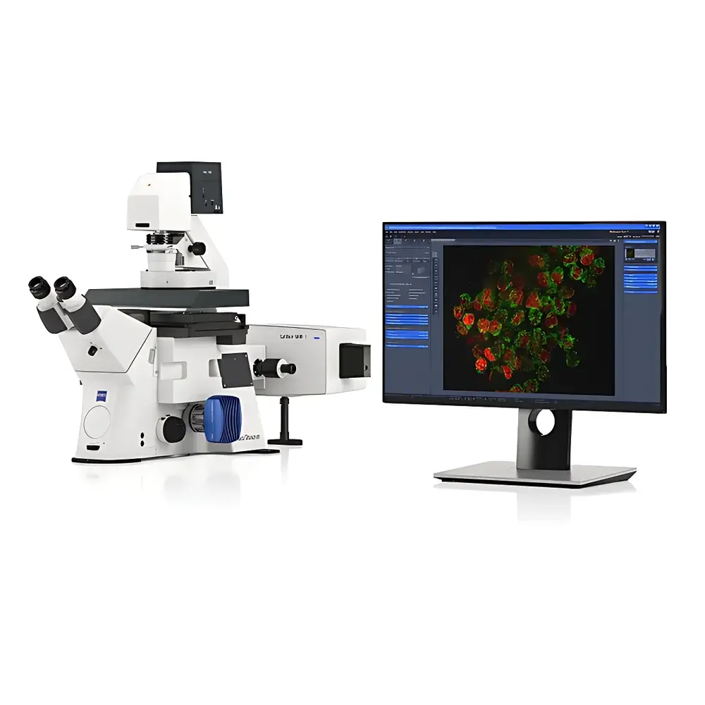
ZEISS Lattice-SIM Series Super-Resolution Microscopy System
| [Brand | ZEISS |
|---|---|
| Origin | Germany |
| Model | Lattice-SIM 3 / Lattice-SIM 5 |
| Imaging Principle | Structured Illumination Microscopy (SIM) with Lattice Light Sheet Integration |
| Resolution (XY) | down to 60 nm (with SIM² reconstruction and Plan-Apochromat 63×/1.40 oil DIC) |
| Optical Sectioning | Yes, via SIM-based optical slicing and TIRF |
| Modalities | SIM, SIM², TIRF, 3D-PALM, SMLM, Leap Mode, Burst Mode |
| Compatibility | Live-cell imaging, fixed specimens, organoids, embryonic tissues, 3D cultures, thick sections |
| Software Platform | ZEN Imaging Software (GLP/GMP-compliant audit trail, FDA 21 CFR Part 11-ready configuration available)] |
Overview
The ZEISS Lattice-SIM Series represents a modular, high-fidelity super-resolution microscopy platform engineered for quantitative structural and dynamic analysis across biological scales—from intact tissue and developing embryos to subcellular protein assemblies and single-molecule localization. Built upon the foundational principles of structured illumination microscopy (SIM), the system integrates lattice-patterned illumination optics with advanced computational reconstruction (SIM²) to surpass the classical Abbe diffraction limit without requiring photoactivatable fluorophores or prolonged acquisition times. Unlike stochastic single-molecule localization techniques (e.g., PALM/STORM), Lattice-SIM delivers deterministic, reproducible resolution enhancement through precisely controlled interference patterns generated by multi-frequency grating sets. This architecture enables simultaneous optimization of spatial resolution, temporal fidelity, and photon budget—critical for low-phototoxicity live-cell imaging and volumetric analysis of optically demanding specimens such as organoids and cleared tissues.
Key Features
- Modular dual-platform design: Lattice-SIM 3 optimized for mesoscopic imaging (10×–40× objectives), Lattice-SIM 5 engineered for nanoscale subcellular dynamics (up to 63×/1.40 oil DIC)
- Five independently tunable spatial frequency gratings per channel, enabling precise matching of illumination pattern pitch to objective NA, emission wavelength, and specimen refractive index
- Dual-mode optical sectioning: SIM Apotome for rapid widefield deconvolution-based sectioning; Lattice-SIM for high-contrast, high-SNR super-resolution sectioning
- Leap Mode and Burst Mode acquisition protocols for synchronized multi-color, multi-plane time-lapse with minimal motion artifact
- Integrated TIRF illumination path for evanescent-field imaging of membrane-proximal events
- Native compatibility with 3D-PALM and single-molecule localization microscopy (SMLM), enabling correlative SIM/SMLM workflows within a single instrument platform
- Plan-Apochromat objective support including 25×/0.8 Imm Corr DIC and 63×/1.40 oil DIC, enabling isotropic resolution down to 120 nm (SIM) and 60 nm (SIM²)
Sample Compatibility & Compliance
The Lattice-SIM Series supports diverse biological preparations without compromise in resolution or viability. Fixed specimens—including resin-embedded ultrathin sections, immunolabeled cryosections, and expansion-microscopy samples—are imaged with sub-100 nm lateral precision. For live applications, the system maintains cellular physiology during extended acquisitions: 3D organoid cultures, zebrafish embryos, Drosophila imaginal discs, and primary neuronal co-cultures are routinely imaged at frame rates up to 10 Hz (depending on FOV and binning) while preserving mitochondrial motility, vesicle trafficking, and cytoskeletal remodeling. All hardware and software components comply with ISO 9001 manufacturing standards. The ZEN software environment supports GLP/GMP-aligned operation with optional 21 CFR Part 11 compliance packages—including electronic signatures, full audit trails, and user-access-tiered permissions—validated for regulated preclinical research environments.
Software & Data Management
Imaging control, reconstruction, and analysis are unified within ZEISS ZEN Imaging Software (v3.6+). SIM² reconstruction employs GPU-accelerated iterative deconvolution with constrained optimization, delivering consistent resolution enhancement independent of fluorophore blinking behavior. Raw data is stored in standardized OME-TIFF format with embedded metadata (objective parameters, laser power, grating position, stage coordinates). Batch processing pipelines support automated drift correction, chromatic aberration registration, and multi-channel alignment. For quantitative colocalization and nanoscale distance mapping, integrated tools include Manders’ coefficients, object-based nearest-neighbor analysis, and FRC (Fourier Ring Correlation) validation metrics. Data export interfaces support HDF5, N5, and BigDataViewer-compatible formats for integration into institutional image informatics infrastructure.
Applications
- Developmental biology: High-speed volumetric imaging of gastrulation, neurulation, and organogenesis in live vertebrate embryos
- Organoid research: Structural phenotyping of epithelial polarity, lumen formation, and intercellular junction maturation
- Neuroscience: Nanoscale mapping of synaptic scaffold proteins (e.g., PSD-95, Bassoon) relative to presynaptic vesicle pools
- Cell biology: Real-time tracking of organelle contact sites (ER–mitochondria, lysosome–autophagosome) with <150 nm spatial discrimination
- Structural virology: Capsid assembly intermediates and envelope glycoprotein clustering in infected cells
- Multiplexed correlative workflows: Sequential SIM → TIRF → SMLM imaging on identical fields-of-view for hierarchical structural validation
FAQ
What distinguishes Lattice-SIM from conventional SIM systems?
Lattice-SIM replaces static binary gratings with dynamically selectable multi-frequency lattice patterns, enabling adaptive illumination geometry that matches both objective point-spread function and fluorophore emission spectra—thereby improving reconstruction fidelity and reducing aliasing artifacts.
Can Lattice-SIM be used for thick-tissue imaging?
Yes—when combined with clearing-compatible objectives (e.g., 25×/0.8 Imm Corr DIC) and adaptive optics modules (optional), the system achieves optical sectioning depths exceeding 100 µm in CLARITY- or CUBIC-processed samples.
Is SIM² reconstruction compatible with third-party fluorophores?
SIM² requires no special labeling chemistry; it functions with standard fluorescent proteins (eGFP, mCherry), organic dyes (Alexa Fluor, ATTO), and quantum dots—provided excitation/emission bands align with available laser lines.
How is phototoxicity managed during long-term live imaging?
The system employs duty-cycle-optimized LED/laser pulsing, real-time intensity feedback control, and low-noise sCMOS detection to maintain photon efficiency below 10 W/cm² at the sample plane—well within established safety thresholds for mammalian cell culture.
Does the platform support automated multi-position time-lapse?
Yes—integrated motorized stages with piezo-driven Z-control and tile-scan stitching enable unattended acquisition across hundreds of positions over 72+ hours, with thermal drift compensation active throughout.


