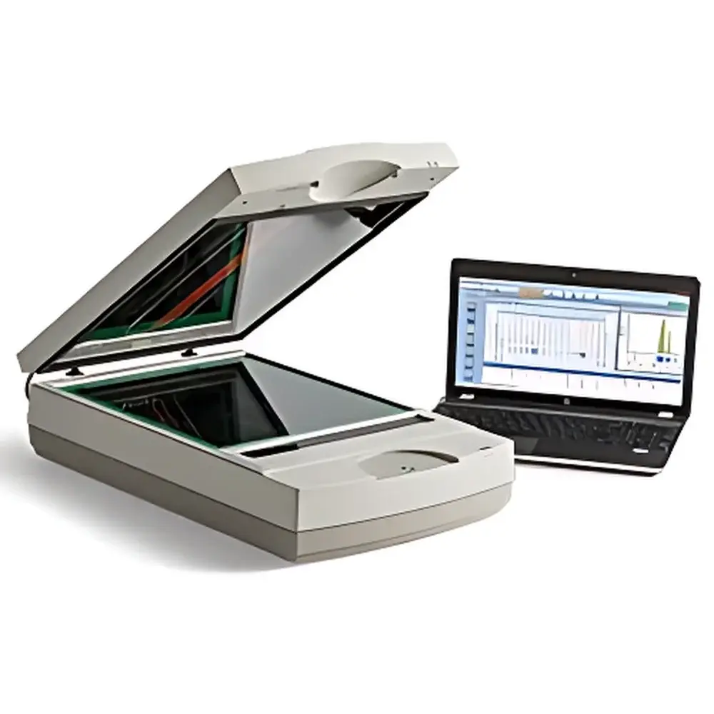
Bio-Rad GS-900 Calibrated Densitometer
| Brand | Bio-Rad |
|---|---|
| Origin | USA |
| Model | GS-900 |
| Instrument Type | Standard Gel Imaging System |
| Bit Depth | 16-bit |
| Optical Density Range | 0.0 to 3.4 OD |
| Illumination Source | LED-based scanning (400–750 nm) |
| Imaging Modes | Transmission and Reflectance |
| Scan Area | 29 × 33 cm |
| Effective Resolution | Adjustable, down to 36.3 µm |
| Image Acquisition Platform | Sealed, humidity-tolerant chamber with adjustable transparent lid |
| Software | Image Lab™ v6.x or later (FDA 21 CFR Part 11 compliant configuration available) |
Overview
The Bio-Rad GS-900 Calibrated Densitometer is a precision optical densitometry system engineered for quantitative analysis of electrophoretic gels, blots, and other planar biosamples under both transmission and reflectance illumination. Unlike conventional CCD-based gel imagers, the GS-900 employs a high-stability LED scanning architecture—rather than a fixed-area camera—to deliver calibrated, reproducible optical density (OD) measurements traceable to NIST-traceable standards. Its core measurement principle relies on photometric scanning across a defined aperture, where light intensity attenuation is converted into linear OD values via logarithmic transformation (OD = log10(I0/I)). This approach ensures minimal spatial distortion, eliminates lens-induced vignetting, and provides inherently uniform illumination across the full 29 × 33 cm imaging field. Designed for routine QC in academic core facilities, biopharma process development labs, and regulatory-compliant environments, the GS-900 meets the metrological requirements for semi-quantitative and quantitative densitometry per ISO/IEC 17025 and ASTM E2893–22 (Standard Guide for Densitometric Analysis of Electrophoretic Gels).
Key Features
- Factory-calibrated optical path with automated self-calibration routine prior to each acquisition—ensures long-term stability of OD values across instrument lifetime
- Adjustable scan resolution (down to 36.3 µm pixel pitch) optimized for either high-throughput screening or high-fidelity band profiling
- 16-bit analog-to-digital conversion (65,536 gray levels), enabling linear response across the full 0.0–3.4 OD dynamic range without signal saturation or digitization loss
- Dual-mode optical design supporting both UV-transparent gel documentation (via blue-light transillumination at 470 nm) and reflective imaging of stained membranes or autoradiographs
- Sealed, temperature-stabilized imaging chamber rated for operation with wet gels up to 10 mm thickness; integrated humidity management prevents condensation during extended acquisitions
- Ergonomic, motorized transparent lid with position-sensing feedback—enables safe, repeatable imaging of variable-thickness samples including thick polyacrylamide gels and multi-layer nitrocellulose membranes
Sample Compatibility & Compliance
The GS-900 accommodates standard electrophoresis formats including mini- and midi-gels (up to 20 × 20 cm), Western blot membranes (PVDF, nitrocellulose), SYBR® Safe-, Coomassie-, and silver-stained gels, as well as ethidium bromide– and GelRed®–stained nucleic acid gels. Its LED spectrum (400–750 nm) avoids UV-induced DNA damage while maintaining sensitivity for common fluorescent dyes. The system complies with IEC 61000-6-3 (EMC emissions) and IEC 61010-1 (safety for laboratory equipment). When configured with audit-trail-enabled Image Lab software, it supports GLP/GMP workflows per FDA 21 CFR Part 11—including electronic signatures, user access controls, and immutable acquisition metadata logging.
Software & Data Management
Image Lab software (v6.1+) provides end-to-end workflow integration—from real-time preview and exposure optimization to band detection, background subtraction, molecular weight estimation, and relative quantification using internal or external standards. All raw scan data are stored in vendor-neutral TIFF format with embedded EXIF metadata (exposure time, lamp intensity, calibration timestamp, OD scaling factor). Batch processing pipelines support ISO/IEC 17025-required uncertainty propagation for replicate measurements. Export options include CSV (for statistical analysis in R or Python), PDF reports with embedded calibration certificates, and XML-based assay definitions compatible with LIMS integration.
Applications
- Quantitative protein expression profiling via SDS-PAGE and Western blot densitometry
- Normalization of qPCR amplicon bands against loading controls (e.g., actin, GAPDH)
- Validation of CRISPR/Cas9 editing efficiency by comparing cleavage band intensities
- Stability-indicating assays for biologics—monitoring aggregate formation in SEC gels
- Regulatory submission support: generation of auditable, calibration-verified densitometry data for IND/BLA dossiers
- Teaching laboratory use: standardized, reproducible gel analysis across multiple student cohorts
FAQ
Does the GS-900 require annual recalibration by a certified service technician?
No—its self-calibration routine, executed before every acquisition using an internal reference tile, maintains traceability to factory-set OD standards. External verification per ISO/IEC 17025 may be performed annually using NIST-traceable neutral density filters.
Can the GS-900 image fluorescent gels without UV exposure?
Yes—the 470 nm blue LED excitation source enables safe, non-UV visualization of SYBR® Safe, SYPRO Ruby, and Deep Purple stains, eliminating ozone generation and photobleaching risks associated with UV transilluminators.
Is the 16-bit data output compatible with third-party analysis tools such as Fiji/ImageJ?
Yes—all acquired images are saved as uncompressed 16-bit TIFF files with documented OD scaling parameters, allowing accurate reprocessing in open-source platforms when metadata-driven linearization is applied.
How does the GS-900 handle uneven sample thickness or warped gels?
The motorized lid’s position-sensing mechanism dynamically adjusts focal plane alignment; combined with diffuse LED illumination, this minimizes shading artifacts and preserves OD linearity across topographic variations up to ±2 mm.
Related Products


