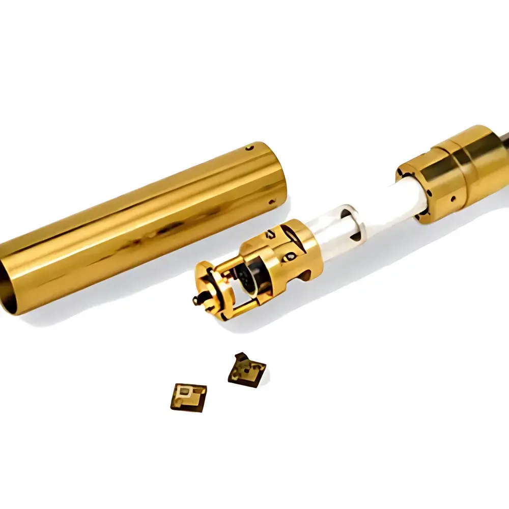
3i LT-SHPM/STM Low-Temperature High-Magnetic-Field Scanning Hall Probe Microscope
| Brand | 3i |
|---|---|
| Origin | Germany |
| Manufacturer Type | Authorized Distributor |
| Origin Category | Imported |
| Model | LT-SHPM/STM |
| Price | Upon Request |
| Max Applied Magnetic Field | ~16 T |
| Operating Temperature Range | 10 mK to 300 K |
| Hall Probe Resolution | ~1 µm |
| STM Resolution | Atomic-scale |
| Scan Ranges (XYZ @ Temp) | 200×200×7.2 µm @ 300 K |
Overview
The 3i LT-SHPM/STM is a cryogenic, high-field scanning probe microscope engineered for quantitative nanoscale magnetometry under extreme thermodynamic conditions. It integrates scanning Hall probe microscopy (SHPM) with complementary scanning tunneling microscopy (STM) and quartz tuning fork (QTF)-based non-contact atomic force microscopy (NC-AFM) in a single ultra-low-vibration platform. Unlike qualitative magnetic imaging techniques such as magnetic force microscopy (MFM), SHPM directly measures the local Hall voltage VH induced by perpendicular magnetic flux density Bz, enabling absolute quantification of magnetic induction (in tesla) with sub-micrometer spatial resolution (~1 µm). The system operates across a continuous temperature range from 10 mK to 300 K and supports static magnetic fields up to ~16 T—enabling studies of quantum phase transitions, vortex dynamics in type-II superconductors, skyrmion lattices, and domain wall pinning in exchange-biased heterostructures.
Key Features
- Quantitative magnetic induction mapping via microfabricated semiconductor Hall probes, calibrated for linear Bz–VH response
- Multi-modal operation: simultaneous or sequential SHPM, STM, and QTF-NC-AFM within identical cryogenic and magnetic field environment
- Three interchangeable scan head configurations optimized for thermal stability and resolution trade-offs: Ultra-Large Area (200×200×7.2 µm at 300 K), Large Area (150×150×7 µm at 300 K), and Standard Area (52×52×4.8 µm at 300 K), each with thermally contracted ranges at 77 K and 4.2 K
- Sub-10 mK base temperature compatibility with dilution refrigerator integration; full compatibility with commercial wet and dry cryostats (e.g., BlueFors, Oxford Instruments, CryoConcept)
- Low-drift piezoelectric positioning with closed-loop capacitive feedback, achieving long-term positional stability <100 pm/h at 10 mK
- Modular design supporting user-defined vacuum feedthroughs, optical access, and RF/DC wiring for in-situ transport measurements
Sample Compatibility & Compliance
The LT-SHPM/STM accommodates conductive, semiconducting, and insulating samples—including thin films, exfoliated 2D materials, epitaxial heterostructures, and bulk single crystals—without requiring conductive coatings or destructive sample preparation. Hall probe operation imposes no topographic constraints, enabling magnetic imaging on rough or patterned surfaces where STM or AFM may fail. All electronics comply with IEC 61000-6-3 (EMC emission standards) and IEC 61000-6-2 (immunity). The control software architecture supports audit trails, electronic signatures, and configurable user roles per FDA 21 CFR Part 11 requirements. Data acquisition protocols are traceable to NIST-traceable voltage and temperature standards, ensuring metrological consistency for ISO/IEC 17025-accredited laboratories.
Software & Data Management
Acquisition and analysis are performed using 3i’s proprietary SHPM-Studio suite, built on a real-time Linux kernel with deterministic latency <50 µs per pixel. The software provides synchronized multi-channel lock-in detection (for Hall voltage, tunneling current, and QTF resonance shift), automated field/temperature ramping with programmable dwell times, and batch-mode correlation of magnetic, topographic, and electronic maps. Raw data are stored in HDF5 format with embedded metadata (field vector, temperature timestamp, probe calibration coefficients, PID parameters). Export modules support MATLAB, Python (via h5py), and IGOR Pro; batch processing pipelines include FFT-based vortex identification, micromagnetic simulation overlays (OOMMF/OOMMF-compatible), and statistical domain analysis compliant with ASTM E112-22 (grain size determination).
Applications
- Direct imaging of Abrikosov vortices and their pinning landscapes in high-Tc cuprates and iron-based superconductors
- Quantitative mapping of stray fields from skyrmions, antiferromagnetic domains, and spin ice monopoles
- In-situ magnetotransport correlation: simultaneous Hall imaging and four-probe resistivity during field sweeps
- Probing proximity-induced magnetism at oxide interfaces (e.g., LaAlO3/SrTiO3) under gate bias
- Validation of micromagnetic simulations against experimentally resolved Bz(x,y) distributions
- Characterization of magnetic dead layers and interfacial anisotropy in multiferroic heterostructures
FAQ
What magnetic field homogeneity is required for quantitative SHPM imaging?
SHPM does not require field uniformity across the entire sample; it measures local Bz. However, field gradients >1 T/mm may induce spurious Hall signals in the probe substrate—system-level field mapping and gradient compensation algorithms are included.
Can the Hall probe be replaced or recalibrated in-house?
Yes. Each Hall sensor is individually characterized and supplied with a NIST-traceable calibration certificate. Replacement probes are pre-aligned and plug-and-play; recalibration routines are integrated into SHPM-Studio and require only a reference solenoid with known field profile.
Is STM operation possible simultaneously with high magnetic fields?
Yes—up to 16 T—with non-magnetic piezoelectric scanners and low-coercivity tungsten tips. Tunneling spectra remain interpretable up to 12 T; above this, Zeeman splitting and orbital effects dominate, which are themselves scientifically valuable for spin-resolved studies.
How is thermal drift compensated during long-duration scans at mK temperatures?
Drift correction uses real-time fiducial tracking of surface landmarks (e.g., step edges or etch pits) combined with predictive thermal model compensation derived from in-situ thermometer arrays mounted on the scanner housing.
Does the system support time-resolved magnetic imaging?
Yes—via external trigger input synchronized to pulsed magnets or RF excitation sources. Minimum frame time is 50 ms at full resolution; sub-pixel dithering enables effective resolution enhancement beyond the probe’s physical footprint.


