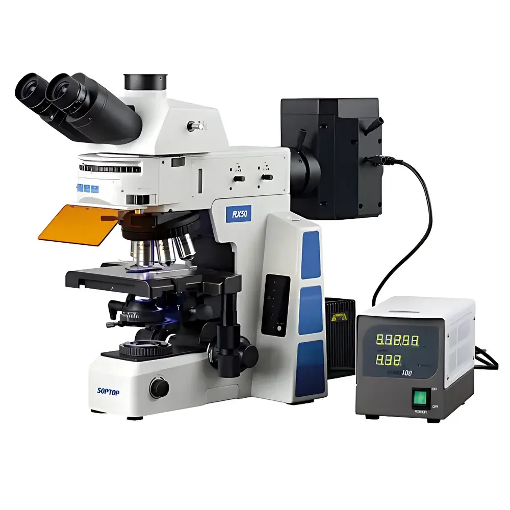
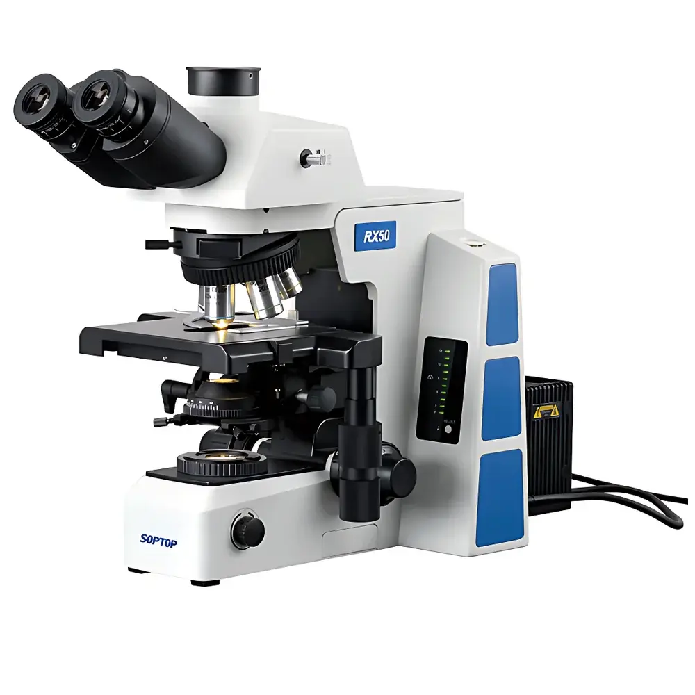
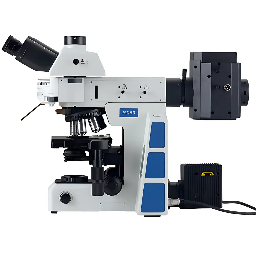
SOPTOP RX50 Research-Grade Upright Fluorescence Microscope
| Brand | SOPTOP |
|---|---|
| Origin | Zhejiang, China |
| Manufacturer Type | Original Equipment Manufacturer (OEM) |
| Product Category | Domestic |
| Model | RX50 |
| Instrument Type | Upright Fluorescence Microscope |
| Excitation Source | 100W High-Pressure Mercury Arc Lamp (optional LED module available) |
| Medical Device Classification | Non-Medical Device |
| Grade | Research-Grade |
| Eyepieces | Widefield Plan Eyepieces PL10X/25mm with Adjustable Diopter, Optional Reticle |
| Objective Lenses | Plan Achromatic (4X, 10X, 20X, 40X, 60X, 100X Oil) |
| Fluorescence Module | Interchangeable Mercury or LED Illumination |
| Transmitted Light Source | 12V/100W Halogen Lamp with Pre-Centered Housing and Continuously Adjustable Intensity |
| Control Mode | Manual |
| Focusing Mechanism | Low-Position Coaxial Coarse/Fine Focus with Adjustable Tension and Programmable Upper Limit Stop |
| Condenser | Universal Condenser (N.A. 0.9) with Built-in Polarizer, Compatible with Phase Annuli, Darkfield Stops, and DIC Prisms |
| Observation Head | Infinity-Corrected Trinocular Hinged Tube (Binocular/Tritocular Configurable) |
| DIC Components | Objective Turret with DIC Slot |
| Stage | 187 × 166 mm Dual-Layer Mechanical Stage with 80 × 55 mm Travel Range, 0.1 mm Positional Resolution, Imported Precision Linear Rails, Bidirectional Drive, Left/Right Hand Configurable, Torque-Adjustable |
| Fluorescence Filter Sets | B1, G1, UV1, B4 |
Overview
The SOPTOP RX50 is an infinity-corrected, research-grade upright fluorescence microscope engineered for high-fidelity cellular and subcellular imaging in life science laboratories. It employs Köhler illumination principles for both transmitted and epi-illumination pathways, ensuring uniform intensity distribution and optimal signal-to-noise ratio across all observation modes—including brightfield, phase contrast, differential interference contrast (DIC), and multi-band fluorescence. Designed around a modular mechanical architecture, the RX50 decouples the optical tube from the main frame, enabling seamless integration of fluorescence, DIC, and polarized light modules without optical realignment. Its rigid aluminum-magnesium alloy chassis minimizes thermal drift and vibration coupling, supporting long-duration time-lapse acquisition and quantitative image analysis workflows.
Key Features
- Modular optical platform with interchangeable illumination arms and condenser systems—enabling rapid reconfiguration between brightfield, phase contrast, DIC, and fluorescence modalities.
- Coaxial coarse/fine focusing mechanism with 25 mm coarse travel and 1 µm fine-step resolution; tension-adjustable knob and programmable upper limit stop ensure repeatable focus positioning for serial sectioning and Z-stack acquisition.
- Universal condenser (N.A. 0.9) with integrated polarizer and accessory slots for phase rings, darkfield stops, and Nomarski prisms—supporting simultaneous optimization of contrast mechanisms without mechanical realignment.
- Trinocular observation head with beam-splitter ratio configurable for simultaneous eyepiece viewing and camera output (e.g., 100:0 / 50:50 / 0:100), compatible with scientific CMOS and sCMOS cameras meeting C-mount or F-mount standards.
- Bidirectional linear-rail stage with 0.1 mm positional repeatability, low-profile design eliminating protruding lateral guides, and torque-adjustable drive for ergonomic operation under extended use conditions.
- Multi-position fluorescence filter turret accommodating up to six excitation/emission/dichroic combinations (B1, G1, UV1, B4 standard); precision-machined indexing ensures zero-image-shift switching during multicolor FISH or co-localization experiments.
Sample Compatibility & Compliance
The RX50 accommodates standard 25 mm diameter glass slides, 35 mm petri dishes, and multi-well plates (6–96-well) using optional stage adapters. Its high-N.A. apochromatic objectives (up to 100X oil immersion, N.A. 1.45) support live-cell imaging with minimal spherical and chromatic aberration across visible and near-UV spectra (350–700 nm). The system complies with ISO 10934-1:2002 (Microscopes — Nomenclature of Microscope Components) and conforms to IEC 61000-6-3:2019 (EMC Emission Standards) and IEC 61000-6-2:2019 (EMC Immunity Requirements). While not classified as a medical device per FDA 21 CFR Part 809 or EU MDR Annex VIII, its optical performance meets ASTM E2867-22 (Standard Guide for Microscope Calibration) and supports GLP-compliant documentation when paired with validated digital imaging software.
Software & Data Management
The RX50 operates independently of proprietary software but integrates natively with third-party acquisition platforms including NIS-Elements (Nikon), ZEN (Zeiss), and open-source solutions such as Micro-Manager 2.0 and Fiji/ImageJ via standard USB 3.0 or GigE Vision interfaces. All hardware controls—including lamp intensity, filter position, focus encoder output, and stage coordinates—are accessible through TTL and RS-232 protocols, enabling full automation in custom-built scripts. Audit trail functionality, user access control, and electronic signature support are achievable when deployed with 21 CFR Part 11–compliant laboratory information management systems (LIMS) or digital microscopy platforms certified for regulated environments.
Applications
- Fluorescence in situ hybridization (FISH) and immunofluorescence (IF) on fixed tissue sections and cultured cells.
- Live-cell dynamics studies using phase contrast and DIC for label-free visualization of organelle motility, mitosis, and cytoskeletal reorganization.
- Quantitative morphometric analysis of neuronal dendritic spines, bacterial colony morphology, or plant stomatal aperture using calibrated plan-apochromatic objectives.
- Multi-modal correlative imaging—combining brightfield histopathology, polarized collagen birefringence, and fluorescence-tagged protein localization on the same specimen.
- Teaching laboratories requiring robust, serviceable instrumentation capable of sustaining daily undergraduate and graduate-level microscopy instruction.
FAQ
Is the RX50 suitable for live-cell imaging?
Yes—the system supports temperature- and CO2-controlled environmental chambers (sold separately), and its LED fluorescence option provides stable, low-heat excitation ideal for prolonged viability assays.
Can the RX50 be upgraded to automated focusing or motorized stage control?
While the base model is manually operated, SOPTOP offers OEM-compatible motorized Z-drive and XY-stage modules with closed-loop encoders, controllable via standard ASCOM or Micro-Manager APIs.
What fluorescence filter sets are included by default?
The standard configuration includes B1 (DAPI), G1 (FITC), UV1 (UV-excited dyes), and B4 (broadband blue excitation) sets; additional sets (e.g., TRITC, Cy5, mCherry) are available as accessories.
Does the RX50 meet regulatory requirements for clinical diagnostics?
No—it is designated for research use only (RUO) and does not carry CE-IVD, FDA 510(k), or NMPA Class II registration for diagnostic applications.
How is optical alignment maintained during modality switching?
All critical optical paths—including condenser height, objective back focal plane, and fluorescence dichroic alignment—are factory pre-centered and mechanically locked; no user recalibration is required when swapping between phase rings or DIC prisms.


