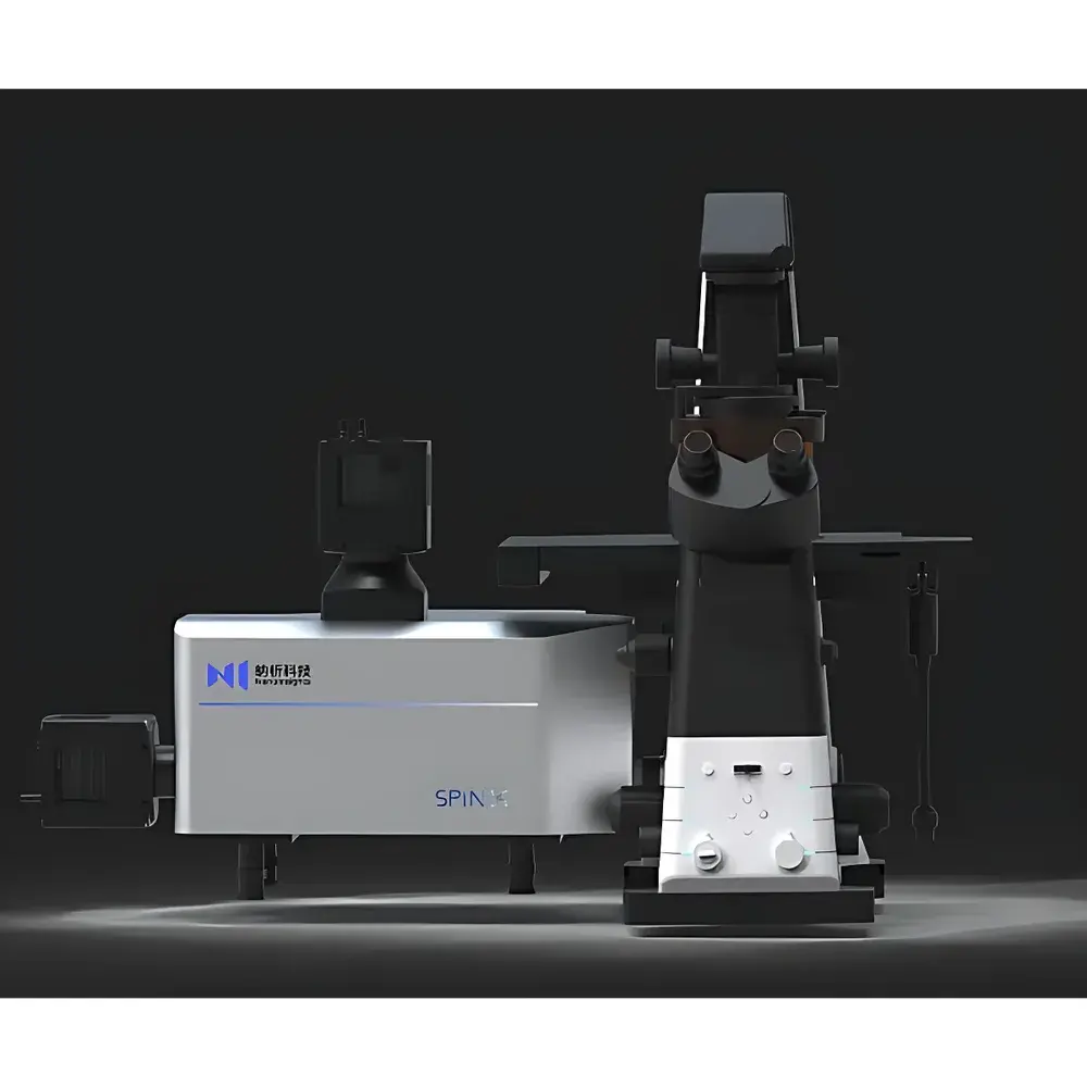
Nanolnsights SpinX Laser Spinning-Disk Confocal Microscope
| Brand | Nanolnsights |
|---|---|
| Origin | Beijing, China |
| Manufacturer Type | Authorized Distributor |
| Origin Category | Domestic (China-made) |
| Model | SpinX |
| Pricing | Available Upon Request |
| Resolution | X-Y = 140 nm, Z = 450 nm |
| Excitation Wavelength Range | 400–750 nm |
| Detection Channels | Fluorescence + Brightfield |
| Fluorescence Detector | Scientific CMOS (sCMOS) Camera |
| Objective Lenses | 10×, 20×, 40×, 60×, 100× (oil/water immersion compatible) |
| Microscope Host | Fully Automated Motorized Platform |
| Illumination Source | Metal Halide Lamp / High-Power LED |
| Control Software | Dual-Mode Intelligent Suite (Beginner & Advanced Modes) |
| Vibration Isolation Table | 1.2 × 1.2 m Dedicated Anti-Vibration Platform |
| XY Stage Drive | Precision Motorized Linear Actuation |
Overview
The Nanolnsights SpinX Laser Spinning-Disk Confocal Microscope is a high-speed, high-fidelity optical imaging platform engineered for quantitative live-cell and 3D tissue microscopy. Built upon the physical principle of Nipkow disk-based confocal illumination, the SpinX employs a precisely aligned, high-rotational-speed micro-perforated disk to achieve parallel multi-point scanning—enabling true optical sectioning without point-scanning latency. Unlike laser-scanning confocal systems, this architecture delivers inherent speed advantages while maintaining diffraction-limited resolution and minimal phototoxic load. With a maximum disk rotation speed of 7500 rpm and frame acquisition rates up to 1440 fps at full sensor resolution, the SpinX is optimized for capturing rapid subcellular dynamics—including vesicle trafficking, calcium wave propagation, mitotic spindle assembly, and organelle motility—in physiologically relevant timeframes.
Key Features
- Spinning-disk confocal architecture with dual-mode excitation: selectable metal halide lamp or high-stability LED sources across 400–750 nm for broad-spectrum fluorophore compatibility (e.g., DAPI, FITC, TRITC, Cy5, Alexa Fluor series)
- High-sensitivity sCMOS detection system offering >82% quantum efficiency, 1.0 e⁻ RMS read noise, and 16-bit dynamic range—critical for low-light, long-duration time-lapse experiments
- Fully motorized microscope host featuring integrated autofocus, automated filter turret, objective revolver, and precision XYZ stage control with sub-micron repeatability
- Dual-path optical design supporting simultaneous brightfield and fluorescence acquisition—enabling correlative morphology-functional analysis without real-time channel switching
- Intelligent software suite with two operational modes: “QuickStart” for standardized protocols and “Expert Mode” granting granular control over exposure timing, z-stack step size, ROI selection, and hardware synchronization
- Dedicated 1.2 × 1.2 m active vibration isolation table, compliant with ISO 20816-1 Class A specifications for optical microscopy environments
Sample Compatibility & Compliance
The SpinX accommodates diverse biological specimens including adherent and suspension mammalian cells, primary neurons, organoids, spheroids, zebrafish embryos (up to 72 hpf), C. elegans, Drosophila tissues, and formalin-fixed paraffin-embedded (FFPE) or cryosectioned tissue slides (up to 2 mm thickness). All optical components meet RoHS Directive 2011/65/EU standards. The system supports GLP-compliant workflows through audit-trail-enabled software logging (user actions, parameter changes, timestamped acquisitions), and is compatible with 21 CFR Part 11–ready data archiving when deployed with validated network storage infrastructure. Objective lenses are certified to ISO 8578 (microscope objective labeling) and JIS B 7151 (numerical aperture tolerance).
Software & Data Management
The SpinX control software is built on a modular, plugin-architected framework compliant with Open Microscopy Environment (OME) standards. Acquired datasets are natively saved in OME-TIFF format with embedded metadata (objective NA, exposure time, z-step, laser power, detector gain). Integrated computational imaging modules include GPU-accelerated Richardson-Lucy deconvolution, non-local means denoising, and deep learning–based enhancement using a pre-trained convolutional neural network (CNN) trained on >50,000 manually annotated confocal image pairs. This AI module demonstrates ≥10× SNR improvement and ≥30% effective resolution gain (measured via Fourier ring correlation, FRC) without increasing illumination dose—validated against NIST-traceable USAF 1951 resolution targets. Raw and processed data are exportable to Fiji/ImageJ, Imaris, Bitplane, and MATLAB via standardized APIs.
Applications
- Live-cell dynamics: Real-time tracking of intracellular transport, membrane receptor internalization, and mitochondrial fission/fusion cycles under physiological CO₂ and temperature control
- 3D culture imaging: High-content volumetric reconstruction of tumor spheroids and iPSC-derived organoids with automated z-focus drift correction
- Drug discovery: Multiplexed phenotypic screening across 96-/384-well plates with adaptive focus mapping and well-to-well intensity normalization
- Neuroscience: Synaptic bouton turnover analysis in primary hippocampal cultures using time-lapse ΔF/F₀ quantification
- Pathology research: Whole-slide fluorescence mapping of multiplexed IHC/IF markers on clinical FFPE sections with tile-scan stitching and spectral unmixing support
- Developmental biology: Long-term (≥24 h) imaging of embryonic morphogenesis in transparent model organisms with adaptive illumination dosing
FAQ
What is the maximum usable field of view (FOV) at 100× oil immersion?
At 100× magnification with a 25 mm field number eyepiece-equivalent sensor, the SpinX achieves a native FOV of 0.19 mm² (275 µm × 275 µm) — expandable via motorized tile-scan up to 10 × 10 mm² with sub-pixel registration accuracy.
Does the system support multi-color time-lapse with hardware-triggered channel switching?
Yes—the filter turret and light source drivers are synchronized via TTL triggers; channel switching latency is <12 ms, enabling precise temporal alignment across ≥4 fluorescence channels.
Is the AI-enhancement module validated for regulatory submissions?
The CNN-based enhancement pipeline is documented per ISO/IEC 17025:2017 Annex A.3 for algorithmic data processing; validation reports (including linearity, precision, and robustness testing) are available under NDA for GxP-regulated users.
Can the SpinX be integrated into automated lab environments?
The microscope supports standard SLIM (Simple Laboratory Instrumentation Messaging) protocol over Ethernet and provides RESTful API endpoints for integration with LIMS, robotic liquid handlers, and environmental chamber controllers.
What maintenance intervals are recommended for the spinning disk and optical alignment?
Disk inspection and recalibration are advised every 12 months or after 5,000 hours of operation; optical alignment verification (using reference PSF beads) is performed automatically during startup and logged in the audit trail.


