Artinis CDMAM 3.4 Mammographic Contrast-Detail Phantom and Analysis Software
| Brand | Artinis |
|---|---|
| Origin | Netherlands |
| Model | CDMAM 3.4 |
| Gold Foil Thickness Range | 0.03–2.00 µm (16 logarithmic steps) |
| Gold Disc Diameter Range | 0.06–2.0 mm (16 logarithmic steps) |
| PMMA Cover Dimensions | 240 × 162 × 3 mm |
| Base Material | 0.5 mm Al (99.5%) |
| PMMA Support Plates | 4 × 10 mm (polished, thickness tolerance ±0.1 mm) |
Overview
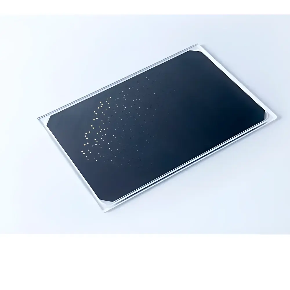
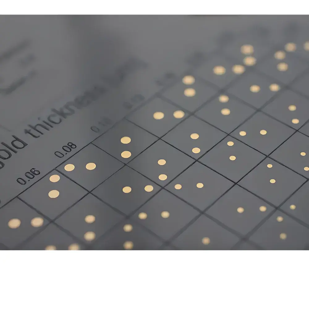
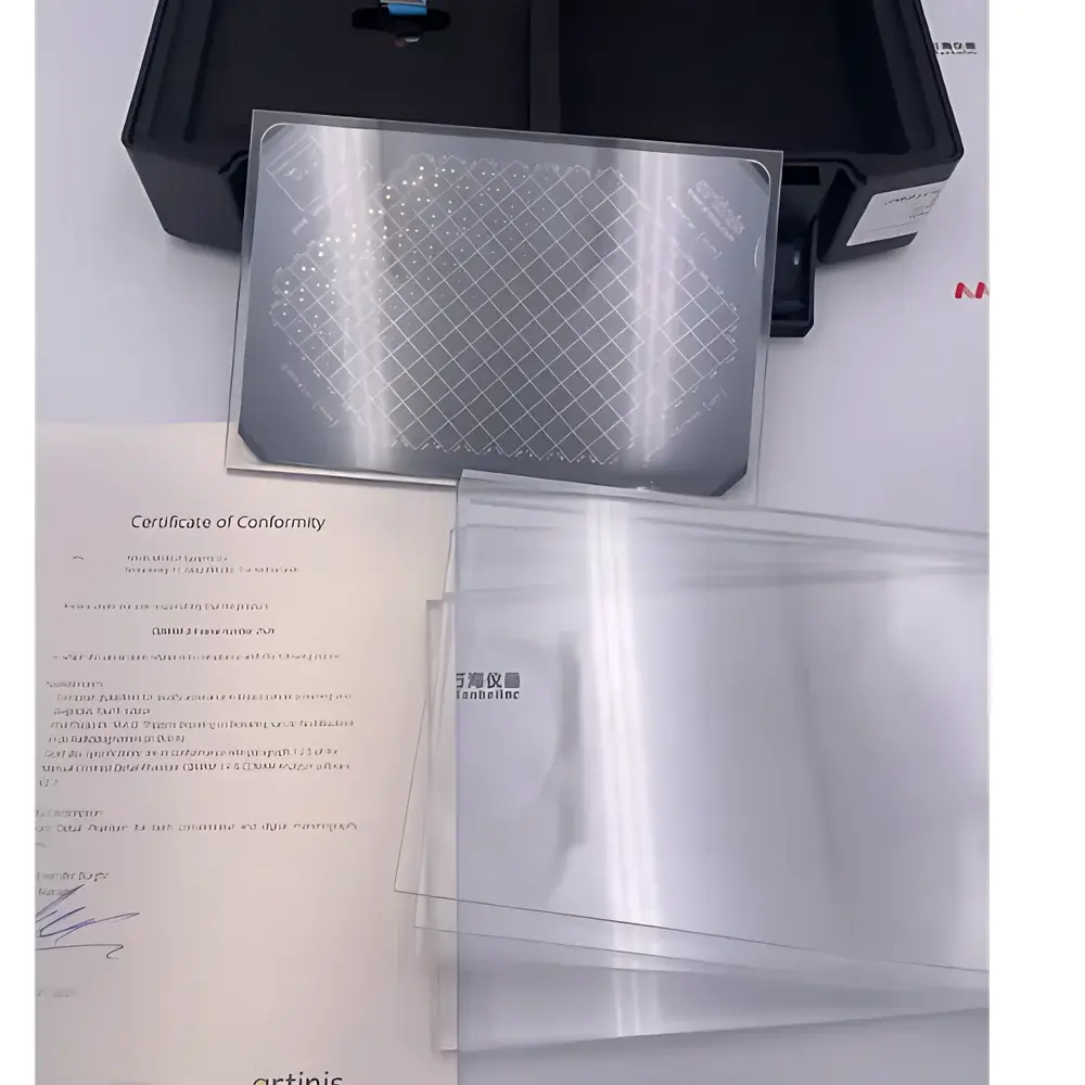
The Artinis CDMAM 3.4 Mammographic Contrast-Detail Phantom is a rigorously standardized test object engineered for quantitative assessment of the low-contrast detectability performance of analog and digital mammography systems. Based on the principles of psychophysical threshold detection, the phantom employs a matrix of gold discs embedded in polymethyl methacrylate (PMMA), systematically varying in both contrast (via controlled gold foil thickness) and size (via discrete disc diameters). This logarithmic arrangement enables precise determination of the observer’s or automated system’s minimum resolvable detail under defined exposure conditions—making it foundational for acceptance testing, routine quality control (QC), and longitudinal performance monitoring in clinical and accreditation settings.
Key Features
- Gold disc array comprising 16 contrast levels (0.03–2.00 µm Au foil thickness) and 16 size levels (0.06–2.0 mm diameter), arranged in a 16 × 16 matrix per quadrant
- Four identical 10 mm-thick PMMA support plates (±0.1 mm dimensional tolerance), ensuring mechanical stability and consistent beam attenuation
- 0.5 mm aluminum base (99.5% purity) providing uniform backscatter conditions and compliance with IEC 61223-3-2 reference geometry
- PMMA cover (240 × 162 × 3 mm) designed to simulate average breast composition and scatter characteristics during clinical acquisition
- Phantom construction adheres strictly to the specifications outlined in the European Guidelines for Quality Assurance in Breast Cancer Screening and Diagnosis (4th Edition) and NHSBSP Equipment Report 0604
Sample Compatibility & Compliance
The CDMAM 3.4 phantom is compatible with all full-field digital mammography (FFDM) systems, computed radiography (CR) mammography units, and film-screen mammography configurations. Its design satisfies mandatory requirements for acceptance and constancy testing under multiple international standards, including IEC 61223-3-2, EN 61223-3-2, and the European Protocol for Physical and Technical Aspects of Digital Breast Tomosynthesis (Version 1.03). The phantom’s geometric and material specifications are traceable to national metrology institutes and validated for use in GLP-compliant QC programs. It supports regulatory audit readiness for facilities undergoing accreditation by EUSOBI, ACR, or national health authorities requiring documented evidence of imaging system performance stability.
Software & Data Management
The CDMAM 3.4 Image Analysis Software (v2.3) implements the CDCOM algorithm developed by the European Reference Organisation for Quality Assured Breast Screening (EUREF). It processes DICOM-compliant mammographic images without proprietary format dependencies. The software performs objective, observer-independent analysis by applying threshold-based detection logic aligned with the European QA Guidelines’ supplementary methodology. Capabilities include batch processing of time-series image sets, generation of contrast-detail (CD) curves, inverse score (IQF) calculation, and tabulated reporting of detected gold disc counts per contrast/size combination. All analyses retain full audit trails—including timestamps, user ID, image metadata, and parameter settings—ensuring compliance with FDA 21 CFR Part 11 requirements for electronic records and signatures when deployed in regulated environments. Reports export to PDF and CSV formats for integration into institutional QC databases.
Applications
- Optimization of exposure parameters (kVp, mAs, AEC settings) across varying breast thicknesses (simulated via PMMA stack configuration)
- Comparative evaluation of image quality between different detector technologies (e.g., amorphous selenium vs. cesium iodide flat panels)
- Validation of automatic exposure control (AEC) consistency and dose efficiency
- Longitudinal tracking of system degradation (e.g., detector lag, gain drift, or focal spot blurring)
- Training and certification of radiographers and medical physicists in standardized QC methodology
- Preparation for external audits and accreditation reviews mandated by national screening programs
FAQ
Is the CDMAM 3.4 phantom suitable for tomosynthesis system evaluation?
Yes—the phantom is explicitly referenced in the European Digital Breast Tomosynthesis QC Protocol (v1.03) for assessing low-contrast detectability in reconstructed slices and projection images.
Does the analysis software require a hardware dongle or network license?
No—licensing is node-locked to the host machine via USB port enumeration; no internet activation or cloud dependency is required.
Can the software process non-DICOM images (e.g., TIFF or JPEG exports from PACS viewers)?
No—only native DICOM files containing complete acquisition metadata (including kVp, mAs, SID, and detector model) are supported to ensure traceable, physics-based analysis.
What is the recommended frequency for CDMAM testing in a clinical mammography program?
Per NHSBSP and EUSOBI guidelines, baseline testing is required at installation and after major service events; routine testing should occur at least quarterly, with monthly checks advised for high-throughput screening units.
How is measurement uncertainty addressed in the software’s detection algorithm?
The CDCOM algorithm incorporates statistical confidence thresholds derived from repeated observer studies; reported IQF values include 95% confidence intervals calculated using bootstrap resampling of detection outcomes across disc subgroups.

")