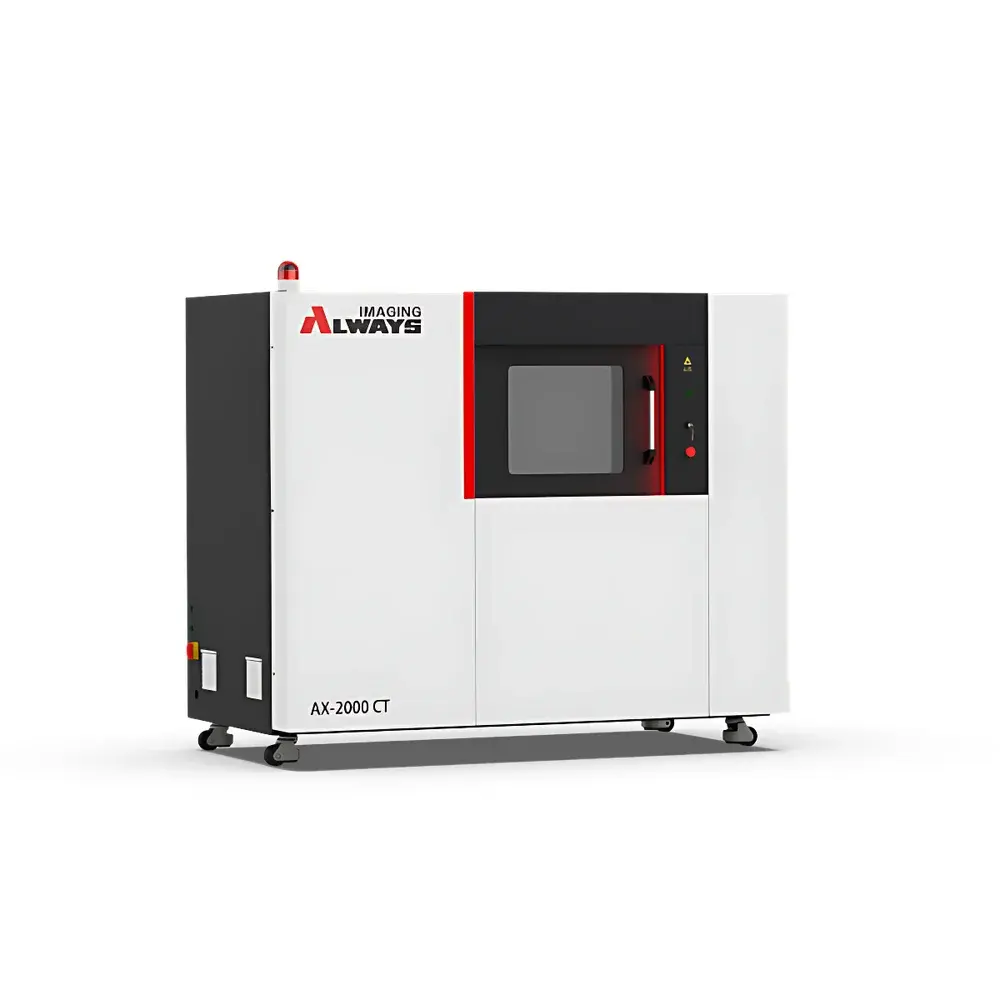
Always Imaging AX-2000CT Compact Industrial Micro-CT System
| Brand | Always Imaging |
|---|---|
| Origin | Zhejiang, China |
| Manufacturer Type | Direct Manufacturer |
| Regional Classification | Domestic (China) |
| Model | AX-2000CT |
| Price Range | USD 210,000 – 350,000 (FOB) |
| Detector Type | Digital Flat-Panel Detector |
| Scan Mode | Translation-Rotation (TR) |
| Spatial Resolution | Up to 0.5 µm (with optional nanofocus source & optical coupling) |
| Density Resolution | <1% contrast sensitivity |
| X-ray Energy | 160 kV / 180 kV (configurable up to 200 kV) |
| Maximum Sample Diameter | 320 mm |
| Maximum Sample Height | 500 mm |
| Maximum Sample Weight | 10 kg (standard), 50 kg (optional rotary stage) |
| System Dimensions (W×D×H) | 1950 × 1050 × 1650 mm |
| System Weight | 1500–2000 kg |
| Radiation Leakage | <1 µSv/h at 1 m |
| X-ray Source | Open-tube transmission target microfocus (1.5 µm focal spot) or nanofocus (0.8 µm focal spot) |
| Tube Current Range | 0.01–0.5 mA |
| Max. Tube Power | 80 W |
| Cooling Method | Air-cooled |
| Detector Pixel Size | 139 µm |
| Detector Array | 3072 × 3072 pixels |
| Field of View (FOV) | 427 × 427 mm |
| Optional Detector | Fiber-optic coupled scintillator with 4×/10×/20× magnification lenses |
Overview
The Always Imaging AX-2000CT is a compact, high-performance industrial micro-computed tomography (micro-CT) system engineered for non-destructive 3D internal inspection and quantitative volumetric analysis. Based on the physical principle of X-ray attenuation tomography—where differential absorption across multiple angular projections enables reconstruction of spatially resolved attenuation coefficients—the AX-2000CT delivers sub-micron structural resolution and high-density contrast fidelity. Its Translation-Rotation (TR) scanning architecture ensures mechanical stability and geometric consistency, minimizing cone-beam artifacts and enabling robust reconstruction of complex geometries without occlusion. Designed for integration into quality control laboratories, R&D facilities, and production-floor environments, the system occupies minimal floor space while maintaining metrological integrity compliant with ISO 12737 (non-destructive testing — industrial computed tomography — qualification of CT systems) and ASTM E1441 (Standard Practice for Computed Tomography (CT) Imaging). The modular design supports field-upgradable X-ray sources and detector configurations, ensuring long-term adaptability to evolving inspection requirements.
Key Features
- Compact footprint (1950 × 1050 × 1650 mm) optimized for constrained laboratory or shop-floor deployment without compromising imaging performance.
- Dual-source capability: Selectable open-tube microfocus (1.5 µm focal spot) or nanofocus (0.8 µm focal spot) X-ray tubes, rated up to 200 kV and 80 W, air-cooled for operational reliability and low maintenance overhead.
- High-fidelity flat-panel detector (3072 × 3072 pixels, 139 µm pitch) providing 427 × 427 mm native FOV; optionally enhanced via fiber-optic coupled scintillator with 4×, 10×, or 20× optical magnification for localized ultra-high-resolution imaging.
- Sub-micron spatial resolution: Achieves ≤0.5 µm effective voxel size under optimal acquisition conditions (nanofocus source + magnified detector mode), validated per ISO/IEC 17025-accredited calibration protocols.
- Density resolution better than 1% contrast sensitivity—enabling reliable discrimination of material phases, porosity distribution, and interfacial defects in multi-material assemblies.
- Comprehensive radiation safety: Lead-shielded cabinet with interlocked access doors; measured leakage dose <1 µSv/h at 1 m distance, meeting IEC 61331-1 and national regulatory requirements for Class II B radiation equipment.
Sample Compatibility & Compliance
The AX-2000CT accommodates diverse sample geometries and material classes within its 320 mm diameter × 500 mm height envelope and 10 kg standard load capacity (upgradable to 50 kg). Validated applications include metallic castings (Al, Mg, Zn alloys), semiconductor packages and bare dies, geological core samples (sandstone, shale, carbonate), polymer composites, additively manufactured lattice structures, battery electrode stacks, and hydrated biological specimens (e.g., bone, plant tissue, insect morphology). All scanning protocols support traceable dimensional metrology per VDI/VDE 2630-1.3 and are compatible with GLP/GMP documentation workflows. System software maintains full audit trails for scan parameters, reconstruction settings, and measurement annotations—supporting FDA 21 CFR Part 11 compliance when deployed with qualified electronic signature modules.
Software & Data Management
Controlled via Always Imaging’s proprietary CT Suite v5.x platform, the AX-2000CT integrates acquisition, reconstruction (FDK, iterative SART, and GPU-accelerated MBIR), segmentation, quantification (porosity, wall thickness, defect sizing), and reporting in a single GUI. Raw projection data is stored in DICOM-CT and HDF5 formats; reconstructed volumes export as 16-bit TIFF stacks or NRRD files for third-party analysis (Avizo, Dragonfly, VGStudio MAX). Batch processing pipelines support automated QA checks against reference standards (e.g., NIST SRM 2088). Data provenance—including source voltage, current, exposure time, rotation step, detector gain, and reconstruction kernel—is embedded in metadata and preserved through all downstream processing stages.
Applications
- Foundry & casting: Detection of shrinkage porosity, inclusions, and mold-filling anomalies in aluminum die-cast components.
- Electronics: Void analysis in solder joints, wire-bond integrity assessment, and encapsulant delamination mapping in QFN/BGA packages.
- Geosciences: Pore-network characterization, fluid saturation dynamics, and mineral phase segmentation in reservoir rock cores.
- Advanced manufacturing: Validation of internal channel geometry in metal AM parts, powder bed fusion defect classification, and lattice strut thickness uniformity.
- Energy materials: Electrode tortuosity quantification, SEI layer thickness distribution in Li-ion cells, and cathode cracking propagation under cycling stress.
- Life sciences: High-resolution morphometric analysis of trabecular bone architecture, root-soil interface visualization, and developmental staging in small-animal models.
FAQ
What is the minimum detectable feature size under standard operating conditions?
With the standard microfocus source and flat-panel detector, the system achieves ≤1 µm effective spatial resolution; using the nanofocus source with 20× optical coupling, resolution improves to ≤0.5 µm in localized regions of interest.
Does the AX-2000CT support automated dimensional metrology per ISO 15530-3?
Yes—when equipped with certified reference objects (e.g., ball plates, step gauges) and operated in calibrated TR mode, the system supports traceable length, diameter, and GD&T measurements compliant with ISO 15530-3 and VDI/VDE 2630-1.3.
Can raw projection data be exported for third-party reconstruction?
Yes—unprocessed sinograms are exportable in HDF5 format with full metadata, enabling custom reconstruction algorithms (e.g., deep learning-based denoising or phase retrieval extensions).
Is remote operation and monitoring supported?
The system includes secure TLS-encrypted web-based remote access for real-time status monitoring, queue management, and diagnostic logging—compatible with enterprise IT security policies.
What regulatory certifications does the AX-2000CT hold?
CE marking (2014/30/EU EMC Directive, 2014/35/EU LVD Directive), RoHS 2011/65/EU compliance, and China NMPA Class II radiation device registration (registration number available upon request).


