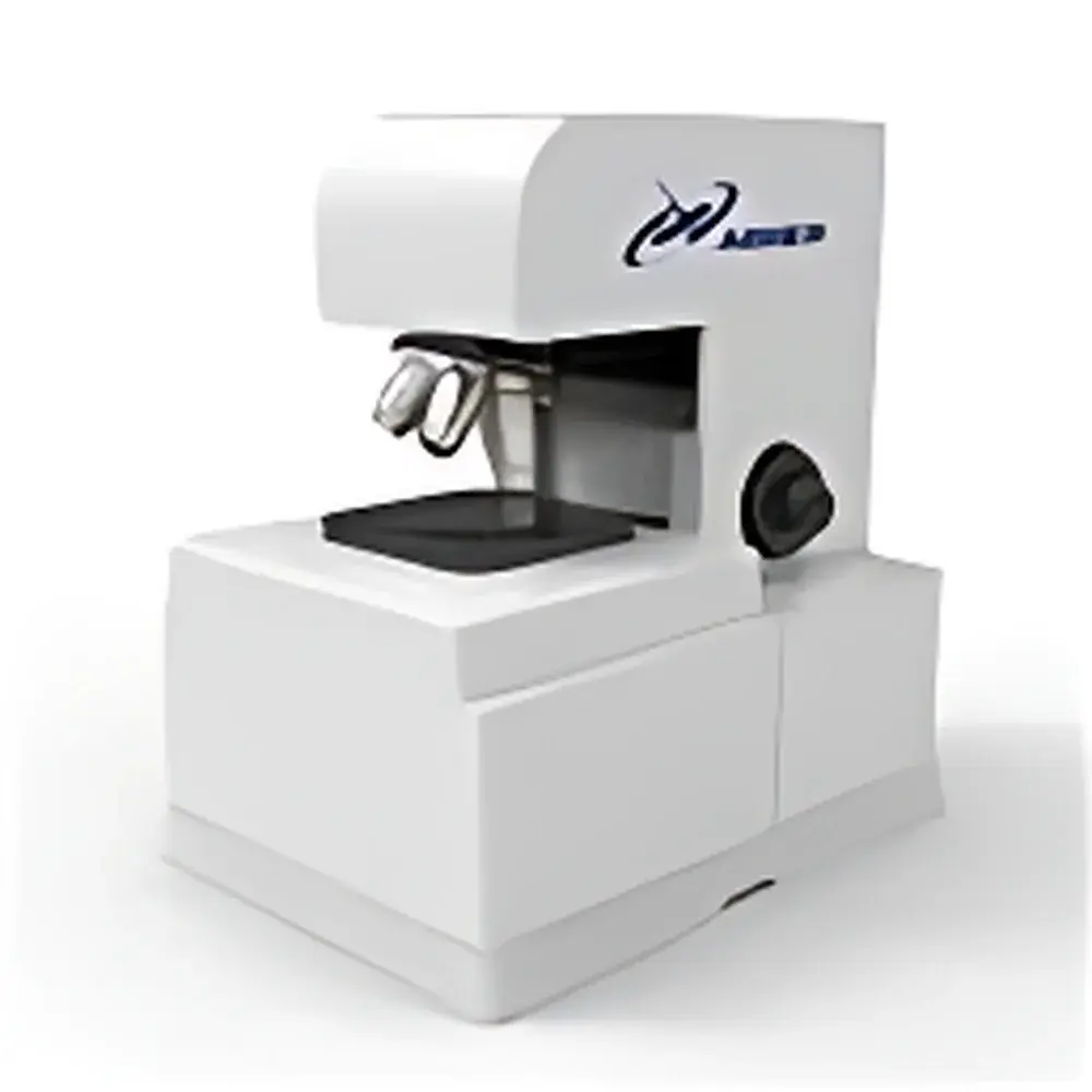
Abner ABN-3DLCM-001 Laser Scanning Confocal Microscope
| Brand | Abner |
|---|---|
| Origin | Jiangsu, China |
| Model | ABN-3DLCM-001 |
| Instrument Type | Point-Scanning Confocal Microscope |
| Resolution (X-Y) | ≤200 nm |
| Resolution (Z) | ≤500 nm |
| Laser Sources | Optional Multi-Wavelength Lasers |
| Detectors | PMT or Multi-Channel Photon Counting Detectors |
| Fluorescence Detection | PMT or Multi-Channel Photon Counting Detectors |
| Scanning Mode | XYZ |
| Objective Lenses | 10×, 20×, 50×, 100× |
| Host Computer | Lenovo Workstation |
| Software & Image Workstation | Fully Integrated Automatic Control |
| XY Stage Control | Motorized and Manual Options |
Overview
The Abner ABN-3DLCM-001 is a high-performance point-scanning laser confocal microscope engineered for nanoscale surface topography and volumetric fluorescence imaging. Based on the fundamental principle of optical sectioning via spatial pinhole rejection, the system eliminates out-of-focus light by aligning the illumination and detection focal planes through a shared confocal aperture—enabling high-contrast, diffraction-limited resolution in both lateral (X-Y) and axial (Z) dimensions. Unlike widefield microscopy, this architecture delivers true optical sectioning capability, allowing sequential acquisition of serial Z-stack images with sub-500 nm axial resolution. The instrument is optimized for non-destructive, label-free or fluorescent 3D morphological characterization across heterogeneous sample classes—including rigid materials, soft biological specimens, thin films, and microfabricated devices—without physical contact or mechanical loading.
Key Features
- Point-scanning architecture with galvanometric mirrors for precise, repeatable XYZ raster scanning and minimal photobleaching in fluorescence mode
- Sub-200 nm lateral resolution and ≤500 nm axial resolution achieved through optimized laser excitation, high-numerical-aperture objectives (10×–100×), and calibrated pinhole alignment
- Modular multi-wavelength laser engine supporting discrete excitation lines (e.g., 405 nm, 488 nm, 561 nm, 640 nm) to accommodate diverse fluorophores and reflectance contrast requirements
- Dual-detection configuration: analog PMT for high-dynamic-range intensity imaging and time-resolved photon counting detectors for quantitative fluorescence lifetime or low-signal applications
- Motorized XYZ stage with closed-loop feedback control, enabling programmable large-area tile scanning, drift-compensated time-lapse acquisition, and reproducible coordinate referencing
- Fully integrated software platform with automated Z-stack acquisition, deconvolution-assisted image restoration, and real-time 3D rendering using GPU-accelerated volume reconstruction algorithms
Sample Compatibility & Compliance
The ABN-3DLCM-001 supports a broad range of sample types without requiring conductive coating or vacuum environments: solid-state materials (metals, ceramics, polymers), semiconductor wafers, MEMS/NEMS devices, fixed or live cells, tissue sections, hydrogels, and optically transparent microfluidic chips. Its non-contact optical measurement modality ensures mechanical integrity preservation—critical for compliant substrates, delicate biospecimens, and post-process inspection of functional microstructures. The system complies with ISO 25178-601 (surface texture—optical areal methods) for quantitative roughness and height parameter extraction. All software operations adhere to GLP-aligned audit trail functionality, including user authentication, timestamped acquisition metadata logging, and immutable raw data storage—facilitating regulatory readiness for QC/QA laboratories operating under ISO/IEC 17025 or FDA 21 CFR Part 11 frameworks.
Software & Data Management
Control, acquisition, and analysis are unified within a dedicated Windows-based application built on a modular architecture. Core modules include: ScanDesigner (for defining region-of-interest, step size, dwell time, Z-interval, and multi-channel synchronization), VoluView (real-time 3D rendering with orthogonal slice navigation, surface rendering, and iso-surface extraction), and QuantifyPro (automated measurement suite for Ra/Rq/Sa/Sq, volume, depth profiles, particle count, and colocalization analysis). Raw image data are stored in open-format TIFF stacks with embedded EXIF metadata; processed results export to CSV, PDF, or structured HDF5 for integration into LIMS or statistical analysis pipelines. Version-controlled software updates include validation documentation per ICH GCP Annex 11 guidelines.
Applications
- Materials Science: Quantitative 3D surface mapping of wear scars, grain boundaries, porosity distribution, and coating uniformity—supporting ASTM E2927 and ISO 4287 compliance
- Semiconductor Metrology: Non-destructive thickness profiling of dielectric layers, trench depth verification, and defect localization on patterned wafers at sub-micron scales
- Life Sciences: High-fidelity 3D reconstruction of nuclear morphology, organelle architecture, and extracellular matrix organization—compatible with standard immunofluorescence and live-cell dyes
- Precision Manufacturing: In-process verification of micro-milled features, laser-ablated textures, and additive-manufactured surface fidelity against CAD reference models
- Academic Research: Teaching platform for optical physics concepts (diffraction limits, Abbe criterion), digital image processing, and computational tomography fundamentals
FAQ
What is the maximum usable field of view at 100× magnification?
At 100× with a 0.95 NA objective, the system achieves an effective field of view of 120 µm × 120 µm per frame, extendable via motorized tiling up to 5 mm × 5 mm with sub-pixel stitching accuracy.
Can the system perform time-lapse 3D imaging of live cells?
Yes—motorized Z-control, low-noise PMT detection, and adjustable laser power enable long-term viability studies; optional environmental chamber integration supports CO₂ and temperature regulation.
Is the software compatible with third-party analysis tools such as Fiji/ImageJ or MATLAB?
All exported TIFF stacks retain full bit-depth and metadata; batch import scripts and API documentation are provided for custom pipeline development.
Does the system support spectral unmixing or FLIM capabilities?
While the base configuration uses discrete-line lasers and intensity-based detection, optional spectral detector modules and TCSPC hardware can be retrofitted for advanced fluorescence characterization.
What maintenance protocols are recommended for long-term stability?
Biannual optical alignment verification, quarterly PMT gain calibration, and annual laser power recalibration are advised—full service logs and SOPs are included with delivery.


