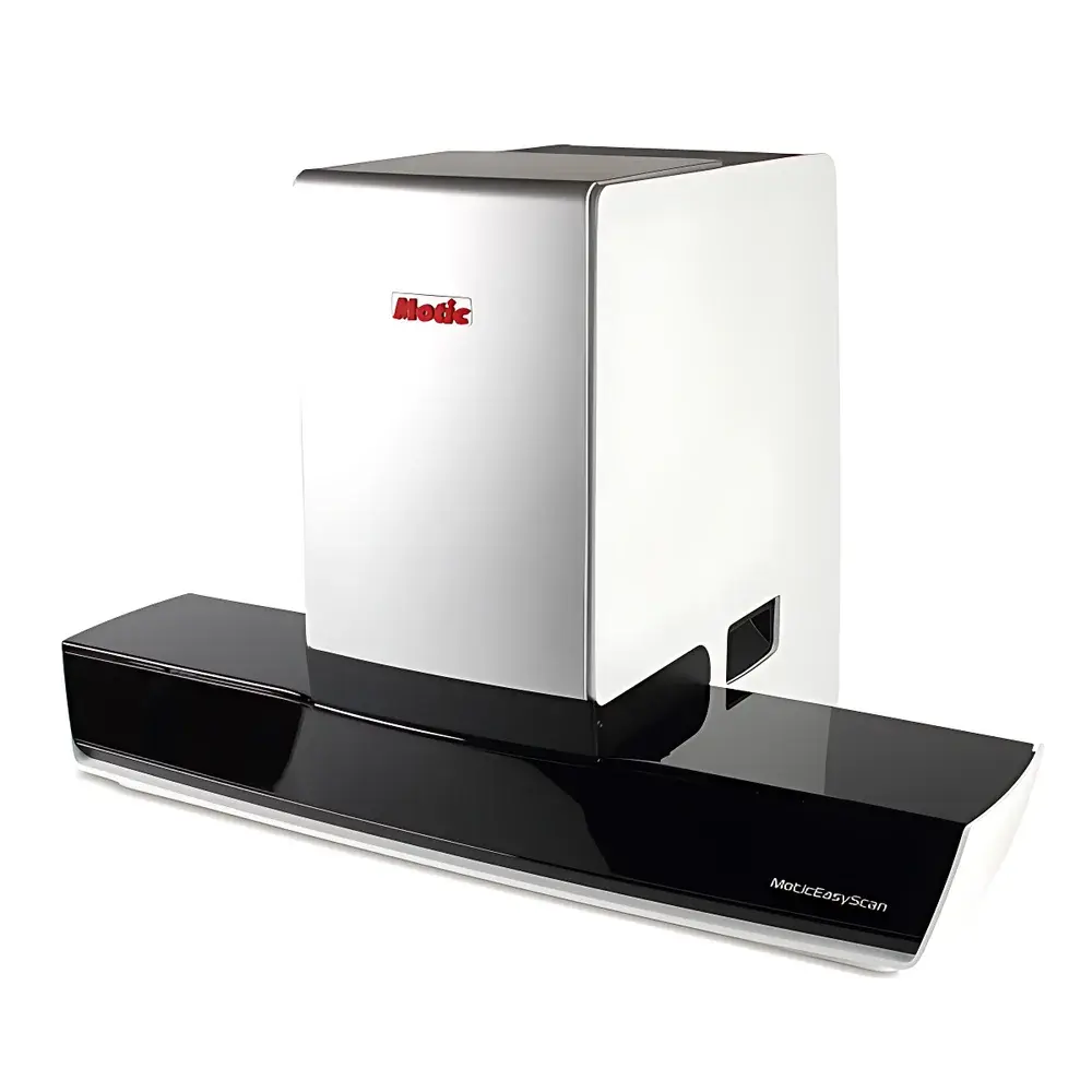
Motic EasyScan 6 Digital Slide Scanner
| Brand | Motic |
|---|---|
| Origin | Fujian, China |
| Manufacturer Type | Authorized Distributor |
| Country of Origin | China |
| Model | EasyScan 6 |
| Pricing | Available Upon Request |
| Objective Lenses | Plan Apochromatic 20×/0.75 and 10×/0.3 |
| Autofocus Technology | Real-time Auto Focus |
| Scan Modes | Standard (Real-time AF), High-Precision AF, Extended Depth of Field (EDF), Z-Stack (Multi-layer Z-direction Scanning) |
| Imaging System | Triple-Camera Architecture |
| Scan Camera | 5 MP, 2/3″ High-Speed CMOS Sensor |
| Pixel Resolution | 0.26 µm/pixel at 40×, 0.52 µm/pixel at 20× |
| Objective Turret | 3-Position |
| Illumination | 10 W LED (25,000 h lifetime) |
| Slide Capacity | 6 slides per batch |
| Slide Dimensions | 76 × 26 mm (tolerance: −0/+1 mm in length, −0/+1 mm in width) |
| Standard Software | EasyScanner, DSAssistant (DSServer optional) |
Overview
The Motic EasyScan 6 Digital Slide Scanner is a compact, high-fidelity whole-slide imaging (WSI) platform engineered for reproducible, high-resolution digitization of conventional glass histopathology and cytology specimens. Based on precision motorized stage translation and synchronized real-time autofocus algorithms, the system captures optical data via a calibrated 5-megapixel 2/3″ CMOS sensor coupled with plan apochromatic objectives (10×/0.3 and 20×/0.75). Each scan generates a seamless, lossless TIFF or SVS-format digital slide—geometrically accurate, photometrically consistent, and compliant with DICOM-SR and OpenSlide interoperability standards. Designed for laboratories transitioning from analog microscopy to digital pathology workflows, the EasyScan 6 delivers clinical-grade image fidelity without requiring enterprise-scale infrastructure. Its modular architecture supports integration into existing PACS, LIS, and EMR environments while maintaining full traceability of acquisition parameters—including objective used, exposure time, white balance, and focus map metadata.
Key Features
- Triple-camera imaging architecture enabling simultaneous brightfield, fluorescence, and transmitted-light channel capture (optional configuration)
- Real-time autofocus engine with sub-micron Z-axis repeatability, ensuring consistent focus across heterogeneous tissue thicknesses
- Four selectable scanning modes: Standard (optimized throughput), High-Precision (sub-pixel stitching alignment), EDF (depth-compensated composite rendering), and Z-Stack (user-defined layer intervals for 3D reconstruction)
- LED illumination with stable color temperature (5700 K ± 150 K) and uniformity >92% across field of view—validated per ISO 15781 for quantitative brightfield imaging
- Motorized 3-position objective turret with mechanical backlash compensation and encoded position feedback for audit-ready objective tracking
- Integrated slide loader accommodating six standard 76 × 26 mm glass slides with automated barcode recognition (ISO/IEC 15415-compliant)
Sample Compatibility & Compliance
The EasyScan 6 accepts unstained, H&E-, IHC-, and special-stained sections mounted on conventional microscope slides (thickness 0.9–1.2 mm). It supports coverslip thicknesses from 0.13–0.17 mm and accommodates common mounting media including DPX, Permount, and aqueous-based alternatives. All scanning protocols adhere to CAP checklist ANP.30710 (Digital Pathology Image Acquisition) and are compatible with FDA-cleared analytical software for primary diagnosis when operated within validated configurations. The system’s firmware logs all acquisition events—including operator ID, timestamp, instrument serial number, and calibration status—in accordance with 21 CFR Part 11 requirements for electronic records and signatures. Routine performance verification follows ASTM E2925-22 (Standard Guide for Validation of Whole Slide Imaging Systems).
Software & Data Management
EasyScan 6 ships with two core applications: EasyScanner (acquisition and quality control interface) and DSAssistant (review, annotation, measurement, and report generation). Both applications support DICOM Structured Reporting (SR) templates for diagnostic consensus and peer review workflows. Optional DSServer enables centralized storage, role-based access control (RBAC), HIPAA-compliant audit trails, and RESTful API integration with third-party AI inference engines (e.g., QuPath, HALO, or custom PyTorch models). All software modules undergo annual penetration testing and maintain ISO/IEC 27001-aligned information security controls. Raw image data is stored in vendor-neutral formats (SVS, NDPI, or TIFF) with embedded EXIF and DICOM header metadata for GLP/GMP traceability.
Applications
- Academic research in histomorphometry, tumor microenvironment quantification, and spatial transcriptomics sample screening
- Clinical pathology departments performing second-opinion consultation, telepathology triage, and digital frozen section review
- Pharmaceutical preclinical labs conducting longitudinal toxicity assessment across rodent and NHP tissue cohorts
- Medical education programs deploying annotated virtual slide libraries aligned with ACGME and FAIMER curriculum standards
- Biobank repositories implementing FAIR-compliant digital specimen indexing and federated search across multi-institutional collections
FAQ
What slide formats does the EasyScan 6 support?
Standard 76 × 26 mm glass slides with thickness between 0.9–1.2 mm; coverslip thickness 0.13–0.17 mm. Custom slide carriers are available upon request for non-standard dimensions.
Is the system compliant with FDA 21 CFR Part 11?
Yes—the acquisition software enforces electronic signature workflows, audit trail logging, and role-based permissions as required under Subpart B of 21 CFR Part 11.
Can the EasyScan 6 be integrated into an existing PACS?
Yes—via DICOM-SR export and HL7 v2.x message routing. Integration documentation and HL7 conformance statements are provided with each installation.
What is the maximum supported magnification for quantitative analysis?
40× objective (0.26 µm/pixel) is validated for morphometric measurements per ISO 13589 and ASTM E2925-22; higher magnifications require external validation by the end user.
Does the system support fluorescence scanning?
Fluorescence capability is available as an optional module with dedicated excitation/emission filter sets and cooled sCMOS sensor upgrade—contact Motic Technical Support for configuration details.


