LA-ICP-MS Imaging Service for Biological Samples
| Brand | Chemlab Pro |
|---|---|
| Origin | Shanghai, China |
| Manufacturer Type | Authorized Distributor |
| Product Category | Domestic |
| Model | LA-ICP-MS Imaging (Biological Applications) |
| Quotation | Upon Request |
| Service Experience | 3–5 Years |
| Sample Types | Animal/Plant Tissue Sections, Fresh Plant Samples, Dried Plant Materials |
| Turnaround Time | 5–10 Business Days |
| Sample Preparation Requirements | Cryosections or resin-embedded sections preferred |
| minimum thickness | 10 µm |
| Sample Mounting | Fresh/dried plant samples must be flat-mounted on conductive stubs |
| max dimensions | 9 cm × 9 cm × 0.8 cm |
| Certified Reference Materials | NIST SRM 1486 (Bone Ash), GBW 07605 (Spinach Leaves), GBW 07607 (Citrus Leaves) |
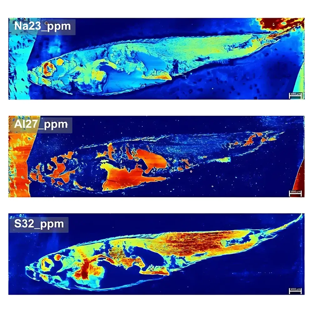
| Zebrafish Imaging | Spatial Resolution: 20 µm |
| Pixel Count | 440,000 |
| Imaging Area | 25.26 mm × 7 mm |
Overview
Laser Ablation Inductively Coupled Plasma Mass Spectrometry (LA-ICP-MS) imaging is a quantitative elemental mapping technique that combines high spatial resolution laser sampling with multi-element detection capability of ICP-TOFMS. This service leverages a fully integrated LA-ICP-TOFMS platform optimized for biological matrix analysis—enabling label-free, simultaneous detection of endogenous metals, metalloids, and exogenous tracers across tissue sections at subcellular-scale resolution. Unlike conventional histological staining or immunoassays, LA-ICP-MS imaging delivers absolute quantification traceable to certified reference materials (CRMs), without reliance on antibody affinity or chromophore stability. The method is particularly suited for metallomics studies, toxicokinetics, drug biodistribution, and developmental biology where elemental co-localization and stoichiometric relationships are critical.
Key Features
- High-fidelity ablation using UV nanosecond laser (e.g., 193 nm ArF excimer or 213 nm Nd:YAG) with precise spot size control (down to 5–20 µm) and programmable raster scanning patterns.
- Time-of-Flight mass spectrometer (TOFMS) enabling full-spectrum acquisition per laser pulse—critical for transient signal capture, isobaric interference correction, and retrospective data reprocessing.
- Quantitative calibration via matrix-matched standards and certified reference materials including NIST SRM 1486 (human bone ash), GBW 07605 (spinach leaves), and GBW 07607 (citrus leaves), ensuring traceability to SI units.
- Robust signal normalization strategies incorporating internal standards (e.g., 13C, 31P, or endogenous 40Ca) to correct for ablation yield heterogeneity and instrumental drift.
- Optimized ion transport interface and low-background vacuum system minimizing polyatomic interferences (e.g., 40Ar16O+, 40Ar12C+) common in biological matrices.
Sample Compatibility & Compliance
This service accepts frozen or resin-embedded tissue sections (minimum thickness: 10 µm); paraffin-embedded sections must undergo complete deparaffinization and ethanol rehydration prior to analysis. Fresh or dried plant samples are accommodated when mounted flat on carbon-coated aluminum stubs with conductive adhesive, conforming to maximum footprint constraints (9 cm × 9 cm × 0.8 cm). All sample handling follows GLP-aligned documentation practices. Data acquisition protocols comply with ASTM E3252–22 (Standard Guide for Elemental Mapping by LA-ICP-MS) and support audit-ready reporting required under ISO/IEC 17025:2017 accreditation frameworks. Raw data files (.raw, .csv) and metadata are retained for ≥5 years in accordance with laboratory retention policies.
Software & Data Management
Data acquisition is performed using vendor-specific control software (e.g., TOFwerk icpTOF Control or Chemlab Pro LA Manager), supporting real-time monitoring of ablation parameters (laser fluence, repetition rate, scan speed) and ion signal intensity. Post-acquisition processing utilizes open-source and commercial tools including Iolite v4 (for time-resolved signal integration and standardization), BioMap (for spatial visualization), and MATLAB-based custom scripts for multivariate statistical analysis (PCA, hierarchical clustering). All reports include pixel-level elemental concentration maps (ng/g or µg/cm²), line profiles, region-of-interest (ROI) statistics, and uncertainty propagation based on CRM uncertainties and counting statistics. Electronic records meet FDA 21 CFR Part 11 requirements for electronic signatures and audit trails where applicable.
Applications
- Metallomic profiling of brain, liver, kidney, and tumor tissues to map dysregulated Fe, Cu, Zn, Mn distributions in neurodegenerative disease models.
- Spatially resolved quantification of platinum-group drugs (e.g., cisplatin, oxaliplatin) and their metabolites in xenograft tumor sections.
- Developmental mapping of essential micronutrients (e.g., Fe, Zn, Se) across zebrafish embryos and larvae—validated at 20 µm resolution over 25.26 mm × 7 mm fields of view (440,000 pixels).
- Phytoremediation studies tracking As, Cd, Pb uptake and compartmentalization in root–shoot systems of hyperaccumulator plants.
- Validation of elemental distribution in CRISPR-edited plant lines exhibiting altered metal transporter expression.
FAQ
What sample preparation is required for optimal LA-ICP-MS imaging results?
For tissue sections: cryosectioning or low-viscosity resin embedding is strongly recommended; paraffin sections must be fully deparaffinized and rehydrated. Minimum section thickness is 10 µm. Samples should be stored at –80 °C prior to sectioning and handled under inert atmosphere if redox-sensitive elements (e.g., Fe2+/Fe3+) are targeted.
Can you perform semi-quantitative analysis without certified reference materials?
No. Quantitative LA-ICP-MS imaging requires matrix-matched calibration standards or CRMs. Semi-quantitative outputs (e.g., relative intensity maps) are not reported unless explicitly requested—and are clearly labeled as non-quantitative in deliverables.
Do you provide raw data and processing scripts?
Yes. Clients receive unprocessed .raw files, calibrated .csv matrices, metadata logs, and documented processing workflows—including Iolite configuration files and BioMap project archives—upon final report delivery.
Related Products
")


