Leica FL400 Blue-Light Fluorescence Module for Neurosurgical Microscopes
| Brand | Leica |
|---|---|
| Origin | Germany |
| Model | FL400 |
| Instrument Type | Integrated Surgical Fluorescence Module |
| Excitation Source | High-Power LED |
| Excitation Range | 380–430 nm (Blue) |
| Emission Detection Range | ≥ 444 nm (Long-Wavelength Blue, Green, Yellow, Red) |
| Regulatory Classification | FDA 510(k)-cleared Class I Device (USA) |
| Compatibility | Leica M530, M620, M720, and other Leica neurosurgical microscopes with TriFluoro-ready architecture |
| Integration | Fully embedded optical path |
| Imaging Output | Full HD (1080p) and optional stereoscopic 3D video recording |
| Software Feature | Mode-Control™ auto-configuration for illumination, exposure, and recording parameters per modality |
Overview
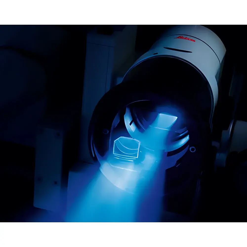
The Leica FL400 Blue-Light Fluorescence Module is an FDA 510(k)-cleared, Class I integrated optical accessory designed exclusively for Leica neurosurgical operating microscopes. Engineered to support intraoperative fluorescence-guided resection, the FL400 operates on the principle of exogenous fluorophore excitation—specifically optimized for 5-aminolevulinic acid (5-ALA)—a prodrug metabolized selectively by malignant glioma cells into protoporphyrin IX (PpIX). Under controlled blue-light excitation (380–430 nm), PpIX emits intense red fluorescence (peak ~635 nm), enabling real-time, high-contrast visualization of tumor tissue against non-fluorescent background brain parenchyma. Unlike standalone fluorescence add-ons, the FL400 is optically and mechanically embedded within the microscope’s light path, preserving native resolution, depth of field, and ergonomic workflow. Its design adheres to ISO 13485 medical device quality management standards and supports compliance with surgical documentation requirements under FDA 21 CFR Part 11 when paired with Leica’s certified recording systems.
Key Features
- Fully integrated blue-light fluorescence pathway—no external beam splitters or alignment procedures required
- High-intensity, thermally stabilized LED excitation source with narrow spectral bandwidth (FWHM < 25 nm) and calibrated irradiance output (≤ 15 mW/cm² at surgical working distance)
- Dual-band interference filter set: excitation filter (380–430 nm) and emission barrier filter (≥ 444 nm long-pass), engineered to maximize signal-to-noise ratio while minimizing photobleaching
- One-touch modality switching between standard white-light mode and FL400 fluorescence mode via ergonomic handle button or footswitch—transition time < 150 ms
- Mode-Control™ software logic automatically adjusts camera gain, exposure time, white balance, and display gamma for optimal rendering in both modalities without manual recalibration
- Native Full HD (1920 × 1080) video output with optional stereoscopic 3D acquisition capability for intraoperative documentation and teaching
- Compatible with Leica’s TriFluoro platform—enables co-registration and sequential activation of FL400 (blue), FL560 (green), and FL800 (near-infrared) modules on a single microscope platform
Sample Compatibility & Compliance
The FL400 module is validated for clinical use with 5-ALA (Gliolan®), administered orally preoperatively at a dose of 20 mg/kg body weight. It is not intended for endogenous fluorescence (e.g., autofluorescence) or non-5-ALA fluorophores without prior validation. The system complies with IEC 60601-1 (medical electrical equipment safety) and IEC 62471 (photobiological safety for lamps and lamp systems). As a Class I device under U.S. FDA regulation, it requires no premarket approval beyond 510(k) clearance (K192577). In the EU, it bears CE marking under Directive 93/42/EEC (now MDR 2017/745) when supplied as part of a certified Leica microscope system. Documentation includes traceable calibration certificates for excitation irradiance and spectral transmission profiles, supporting GLP/GMP-aligned surgical audit trails.
Software & Data Management
FL400 operation is managed through Leica’s LAS X surgical imaging platform, which provides DICOM-compliant image capture, timestamped video logging, and metadata tagging (modality, exposure settings, surgeon ID, case ID). All recordings include synchronized dual-stream output: one channel preserves native white-light reference frames; the second encodes fluorescence-enhanced view with overlay annotation capability. Audit trail functionality meets FDA 21 CFR Part 11 requirements when deployed with Leica’s secure authentication server and electronic signature module. Export formats include MP4 (H.264), AVI (uncompressed), and DICOM-SR for PACS integration. Firmware updates are delivered via encrypted USB key or network-based secure OTA protocol, with version rollback and integrity verification.
Applications
- Intraoperative delineation of high-grade glioma margins during supratentorial craniotomy, improving gross-total resection rates while preserving eloquent cortex
- Real-time assessment of residual tumor burden following initial debulking, reducing repeat surgery incidence
- Supporting fluorescence-guided biopsy targeting in suspected low-grade gliomas where contrast enhancement is absent on preoperative MRI
- Training and simulation environments—FL400’s consistent spectral output enables standardized fluorescence response across cadaveric and phantom models
- Multi-modal correlation studies integrating FL400 data with intraoperative ultrasound or neuronavigation datasets via Leica’s FusionNav interface
FAQ
Is the FL400 compatible with non-Leica microscopes?
No. The FL400 is a proprietary optical module requiring precise mechanical coupling, beam path alignment, and firmware-level integration with Leica’s M530, M620, and M720 neurosurgical platforms.
Does FL400 require special maintenance or recalibration?
The LED light source is rated for > 20,000 hours of operation with negligible spectral drift. Annual performance verification using Leica’s calibrated photometric test kit is recommended but not mandatory per regulatory requirements.
Can FL400 be used with other fluorophores such as ICG or fluorescein?
ICG and sodium fluorescein are not spectrally compatible with FL400’s excitation/emission bandpass configuration. For ICG, Leica FL560 (green excitation) or FL800 (NIR) modules are indicated.
What documentation is provided for regulatory submission support?
Leica supplies a Technical File Summary, Declaration of Conformity, FDA 510(k) summary letter, and filter spectral transmission reports—sufficient for hospital biomedical engineering review and IRB protocol submissions.
Is 3D recording available on all Leica microscope models?
3D capability requires optional stereoscopic camera hardware and is supported only on M530 and M720 platforms with dual-sensor acquisition modules.


