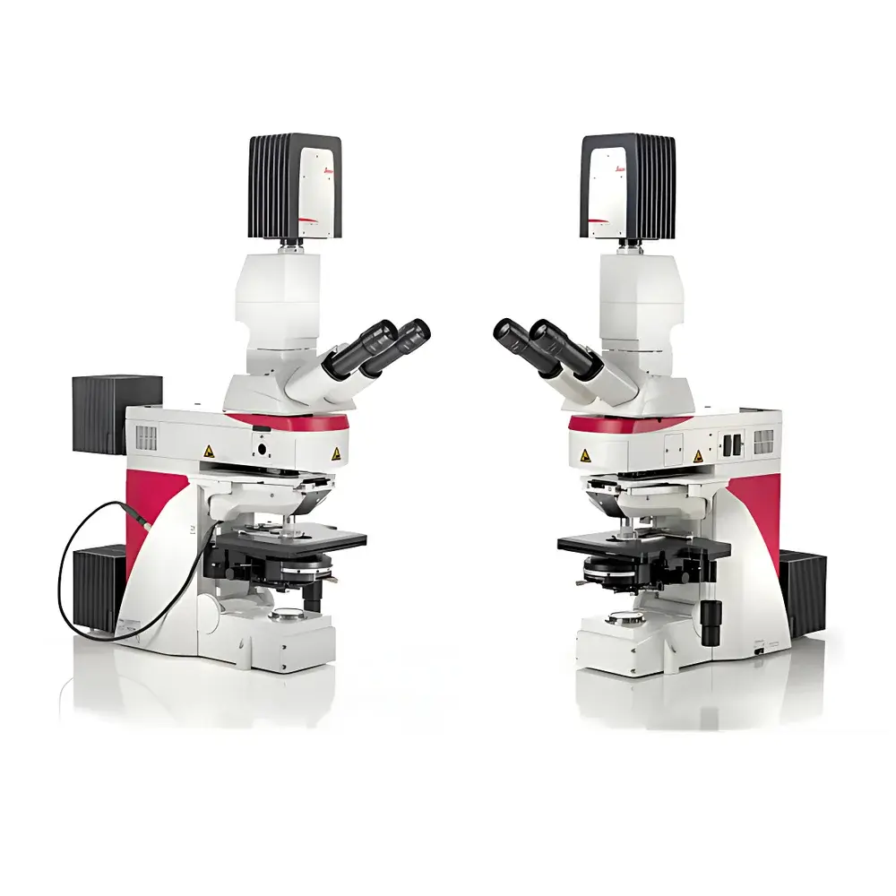
Leica DM6 FS Upright Intelligent Microscope
| Brand | Leica |
|---|---|
| Origin | Germany |
| Model | DM6 FS |
| Type | Upright Fixed-Stage Widefield Microscope |
| Application Focus | Electrophysiology, In Vivo Imaging, Optogenetics, Cryo-CLEM Integration |
| Compliance | Designed for GLP/GMP-adjacent research environments |
Overview
The Leica DM6 FS is an upright, fixed-stage widefield microscope engineered specifically for demanding life science applications where mechanical stability, optical fidelity, and experimental integrity are non-negotiable. Built upon the same rigid, low-vibration mechanical platform as the Leica DM6 B, the DM6 FS eliminates micro-motion during critical manipulations—making it a foundational instrument for patch-clamp electrophysiology, optogenetic stimulation, in vivo tissue imaging, and correlative light-electron microscopy (CLEM) workflows. Its core architecture employs a massive, monolithic base casting and passive damping elements to suppress ambient vibrations below 1 Hz, ensuring sub-micron positional stability during prolonged recordings or laser-based interventions. Unlike conventional motorized upright systems, the DM6 FS integrates fully decoupled electronic actuation: all motorized components—including objective turret, focus drive, filter wheels, and illumination shutters—are independently controllable and can be powered down during sensitive acquisition phases, eliminating electromagnetic interference and thermal drift. This design philosophy directly addresses the stringent requirements of neuroscience laboratories performing simultaneous optical stimulation and electrical recording.
Key Features
- Fixed-stage mechanical architecture with ultra-low vibration transmission (<0.5 µm RMS displacement under standard lab conditions)
- Fully motorized, software-controlled operation via Leica LAS X—no physical interaction required during acquisition
- Independent power gating for all motorized subsystems to eliminate EMI and thermal load during silent acquisition windows
- Expanded working clearance above the stage (≥35 mm vertical access) for unobstructed micromanipulator integration and pipette approach angles up to 41°
- Optimized optical path for near-infrared (NIR) and visible-light compatibility, supporting dual-channel optogenetic stimulation and fluorescence detection
- Dedicated DIC optics using Wollaston prisms and strain-free objectives for high-contrast, high-resolution imaging through thick, scattering specimens (e.g., brain slices ≥300 µm)
- Modular expansion capability for integration with Leica EM Cryo CLEM systems, including cryo-stage adapters and vacuum-compatible optical ports
Sample Compatibility & Compliance
The DM6 FS accommodates standard glass-bottom dishes, multi-well plates, acute brain slices (200–500 µm), explanted organs, and live-animal head-fixed preparations. Its large working distance objectives—including the Leica HC FLUOTAR 25×/0.95 W VISIR (2.5 mm WD, 41° entry angle)—enable simultaneous optical access and electrophysiological probing without collision risk. The system complies with ISO 10934-1 (microscope optical performance standards) and supports traceable calibration protocols aligned with ASTM E2812 for quantitative fluorescence intensity measurement. When deployed with Leica LAS X software configured for audit trail logging, electronic signatures, and secure user roles, the platform meets functional requirements for GLP-compliant documentation and 21 CFR Part 11–aligned data integrity frameworks.
Software & Data Management
Control and acquisition are managed exclusively through Leica LAS X software—a modular, scriptable platform supporting Python-based automation, time-lapse scheduling, multi-dimensional acquisition (XYZTλ), and real-time Z-stack reconstruction. LAS X provides full hardware abstraction: all motors, shutters, and illumination sources are addressable via API calls, enabling seamless synchronization with external devices (e.g., patch-clamp amplifiers, TTL-triggered lasers, or DAQ systems). Raw image data is stored in standardized TIFF or Leica’s proprietary LIF format, both embeddable with EXIF-like metadata (objective ID, exposure, gain, stage coordinates, timestamp). Optional LAS X Security Pack enables role-based access control, electronic signatures, and immutable audit trails—critical for regulated preclinical research environments.
Applications
- Electrophysiology: Stable platform for whole-cell patch-clamp on neurons in acute slices or cultured preparations; minimal stage drift preserves seal integrity over >30-minute recordings
- Optogenetics: Precise co-localization of 473 nm/594 nm stimulation beams with NIR imaging channels; synchronized shutter control prevents phototoxicity during baseline periods
- In Vivo Imaging: Compatible with custom head-plate mounts and cranial window configurations; large vertical access allows angled objective positioning for thalamic or cortical layer targeting
- Cryo-CLEM: Native integration point for Leica EM Cryo-Transfer systems; optical maps generated at room temperature are spatially registered to cryo-EM grids with sub-100 nm correlation accuracy
- Developmental Biology: Long-term time-lapse of zebrafish or Drosophila embryos under physiological conditions, leveraging low-heat LED illumination and automated focus maintenance
FAQ
Is the DM6 FS compatible with third-party electrophysiology rigs?
Yes—the microscope features standardized TTL and analog I/O ports, and LAS X supports LabVIEW and MATLAB interoperability via TCP/IP and COM interfaces.
Can the system be used inside a Faraday cage?
All motor drivers and controllers are housed within the base unit; external cabling uses shielded, filtered connectors—enabling full functionality inside grounded electromagnetic isolation enclosures.
Does Leica provide validation documentation for GxP environments?
While the DM6 FS itself is not a medical device, Leica offers IQ/OQ documentation templates and supports vendor-assisted installation qualification for labs operating under GLP or ISO 17025 frameworks.
What immersion media are supported by the VISIR objective?
The HC FLUOTAR 25×/0.95 W VISIR is optimized for water, saline, and agarose-based immersion; it is not designed for oil or glycerol immersion.
How is thermal stability maintained during extended acquisitions?
The base casting incorporates thermally symmetric design and passive heat-sinking; active cooling is omitted to avoid airflow-induced vibration—ambient temperature fluctuations ≤±0.5°C/h are recommended for sub-pixel registration stability.


