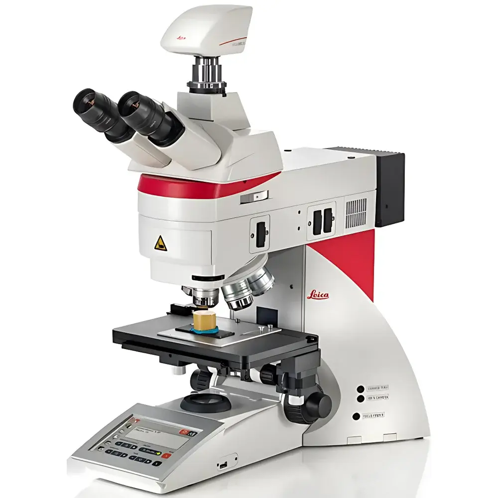
Leica DM6 M LIBS Microstructural Composition Analysis System
| Brand | Leica |
|---|---|
| Origin | Germany |
| Model | DM6 M LIBS |
| Application | Integrated Optical Microscopy + Laser-Induced Breakdown Spectroscopy (LIBS) for Real-Time Elemental Mapping |
| Sample Preparation | None Required |
| Analysis Speed | <1 Second per Spot |
| Spatial Resolution | ~5–10 µm (laser spot size dependent) |
| Spectral Range | 200–900 nm |
| Detection Capability | Qualitative & Semi-Quantitative Elemental Identification (Z ≥ 5, i.e., Boron and above) |
| Compliance | CE Marked, ISO 9001 Manufacturing Environment, Designed for GLP/GMP-Adjacent R&D and QC Environments |
Overview
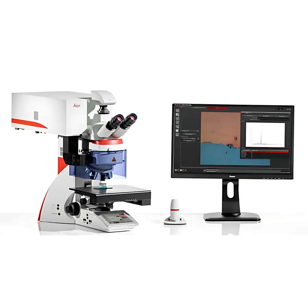
The Leica DM6 M LIBS Microstructural Composition Analysis System is an integrated optical microscopy and laser-induced breakdown spectroscopy (LIBS) platform engineered for concurrent morphological observation and rapid elemental characterization at the microscale. Unlike conventional workflows requiring sequential optical imaging (e.g., brightfield, DIC, or polarized light) followed by vacuum-based electron microscopy with energy-dispersive X-ray spectroscopy (SEM/EDS), the DM6 M LIBS performs both functions in a single instrument, under ambient atmospheric conditions, without sample coating, sectioning, or conductive treatment. Its core principle relies on focused nanosecond-pulsed laser ablation to generate transient microplasmas on the sample surface; emitted atomic/ionic line spectra are collected via high-resolution Czerny–Turner spectrometer and matched against reference spectral libraries for elemental identification. This enables real-time, location-specific chemical mapping directly within the field of view—preserving spatial context between structure and composition.
Key Features
- Seamless integration of research-grade upright microscope optics (Leica DM6 M) with a collinear, fiber-coupled LIBS module—no beam path compromise or image distortion.
- Automated co-registration: The system maintains sub-micron alignment between optical imaging coordinates and LIBS analysis position—eliminating ROI relocation errors common in hybrid SEM-LIBS or stage-transfer setups.
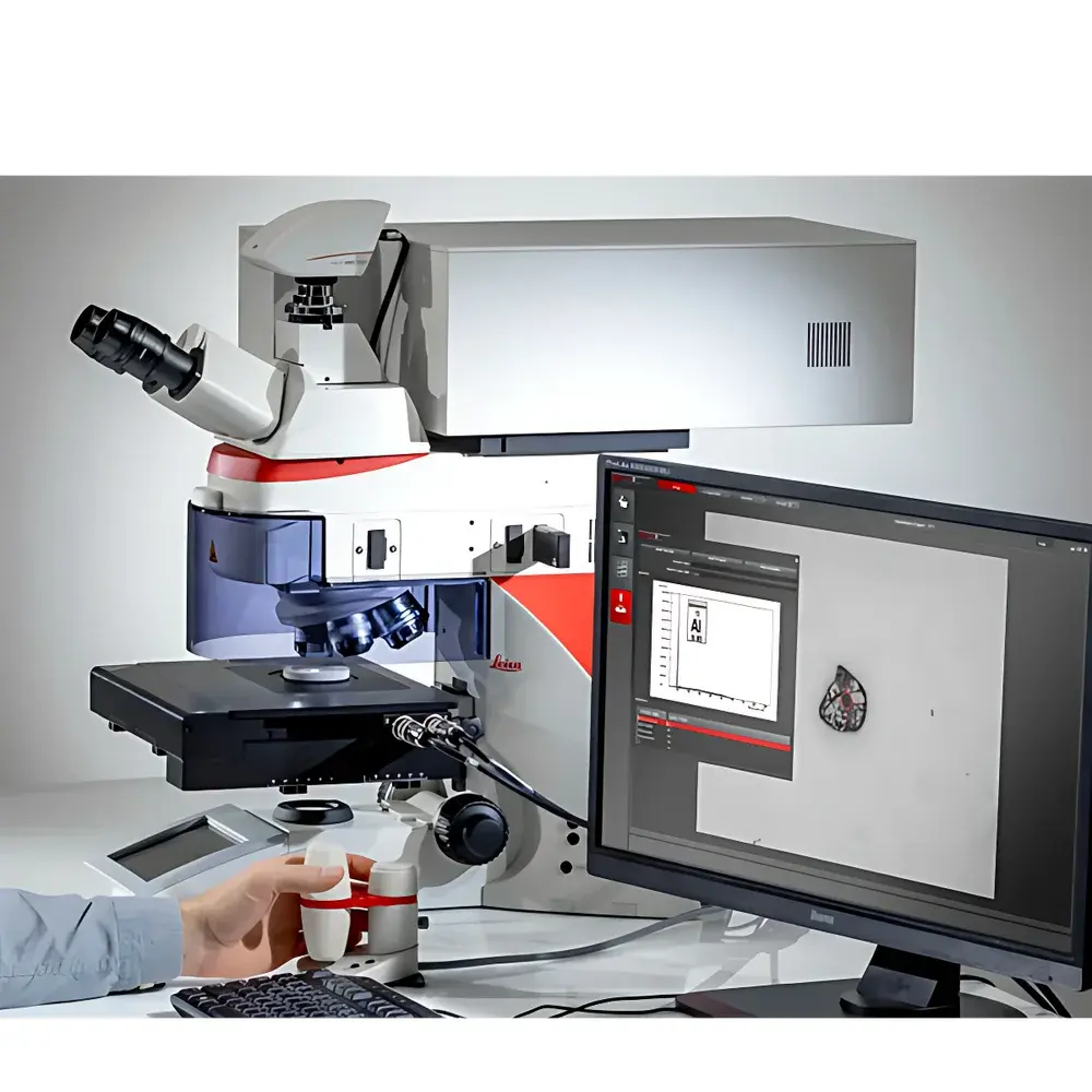
- One-click elemental interrogation: Select any region visible through eyepiece or digital camera; initiate LIBS acquisition with a single software command—no manual focusing, plasma optimization, or spectral calibration required between measurements.
- Zero-sample-prep operation: Compatible with bulk metals, ceramics, polymers, geological specimens, filters, and coated substrates—regardless of conductivity, topography, or volatility.
- Multi-contrast imaging support: Leica’s HCX PL Fluotar objectives (1.25×–100×) combined with transmitted light (brightfield, phase contrast, polarization) and reflected light (DIC, fluorescence) ensure high-fidelity structural context prior to chemical interrogation.
- Modular upgrade path: Existing Leica DM6000 M or DM6 M microscope users can retrofit the LIBS module without replacing the base system—reducing capital expenditure and preserving legacy optical calibration records.
Sample Compatibility & Compliance
The DM6 M LIBS accommodates heterogeneous, irregular, and non-conductive samples—including as-received industrial filters, fractured metallographic sections, uncoated ceramic tiles, and embedded particulates—without vacuum chamber constraints or charge dissipation requirements. It complies with CE marking directives for laboratory equipment safety (2014/30/EU EMC, 2014/35/EU LVD) and is manufactured under ISO 9001-certified processes. While not a regulated medical device, its design supports audit-ready workflows aligned with GLP principles: full traceability of acquisition parameters (laser pulse energy, gate delay, integration time), user-defined spectral library versioning, and timestamped metadata export. For regulated environments, optional audit trail logging (user ID, session start/end, analysis trigger events) meets foundational expectations for FDA 21 CFR Part 11–aligned documentation—though formal validation remains site-specific.
Software & Data Management
Leica Application Suite X (LAS X) serves as the unified control interface, providing synchronized microscope navigation, live imaging, LIBS acquisition, and spectral interpretation in one environment. Each LIBS spectrum is automatically annotated with XY-stage coordinates, objective magnification, illumination mode, and laser settings. Elemental maps are generated via peak-intensity ratio algorithms applied to selected emission lines (e.g., Fe I 371.99 nm, Al I 396.15 nm); false-color overlays are superimposed directly onto optical images. Spectral libraries include NIST Atomic Spectra Database (ASD) references and allow user-defined additions—enabling iterative refinement for alloy-specific or contaminant-targeted detection. All raw spectra, processed maps, and metadata export to open formats (.csv, .tif, .spc), ensuring interoperability with third-party chemometric tools (e.g., MATLAB, Python SciPy) for advanced multivariate analysis.
Applications
- Contamination root-cause analysis: Rapid identification of metallic, siliceous, or organic particles on filtration media—correlating morphology (e.g., spherical vs. angular) with chemistry (e.g., Cu vs. SiO₂) in one workflow.
- Coating and layered material characterization: Depth-profiling via sequential laser pulses to resolve compositional gradients across paint layers, PVD coatings, or oxide scales—without cross-sectioning.
- Mineralogical and metallurgical phase assessment: Discrimination of intermetallics, inclusions, or gangue minerals based on combined structural contrast and elemental signatures—supporting ASTM E112 grain size or E3 standard practices.
- Failure analysis triage: Pre-screening of fracture surfaces or corrosion sites to determine whether further SEM/EDS or TEM investigation is warranted—reducing backlog by >90% in routine QA labs.
- Cleanliness verification (ISO 16232, VDA 19): Paired with Leica Cleanliness Expert software, it automates particle counting, sizing, and classification—including elemental attribution—to meet automotive and aerospace particulate standards.
FAQ
Does DM6 M LIBS require vacuum or conductive coating?
No. All analyses occur at ambient pressure and temperature. Non-conductive samples (e.g., polymers, oxides) are analyzed directly without sputter coating.
Can it quantify elemental concentrations?
It provides semi-quantitative results based on relative peak intensities and matrix-matched calibration standards; absolute quantification requires certified reference materials and empirical calibration curves.
What is the lateral resolution of LIBS mapping?
Spatial resolution is governed by laser spot size (typically 5–10 µm with standard optics) and plasma expansion—making it suitable for microstructural features ≥10 µm.
Is depth profiling destructive?
Yes—LIBS is inherently ablative. Each pulse removes ~10–100 nm of material; cumulative ablation enables controlled layer-by-layer analysis but alters the local surface.
How does it compare to SEM/EDS in detection limits?
DM6 M LIBS offers faster screening and superior light-element sensitivity (B, C, N, O), whereas SEM/EDS provides lower detection limits (<0.1 wt%) for heavier elements and higher spatial resolution for nano-scale features.


