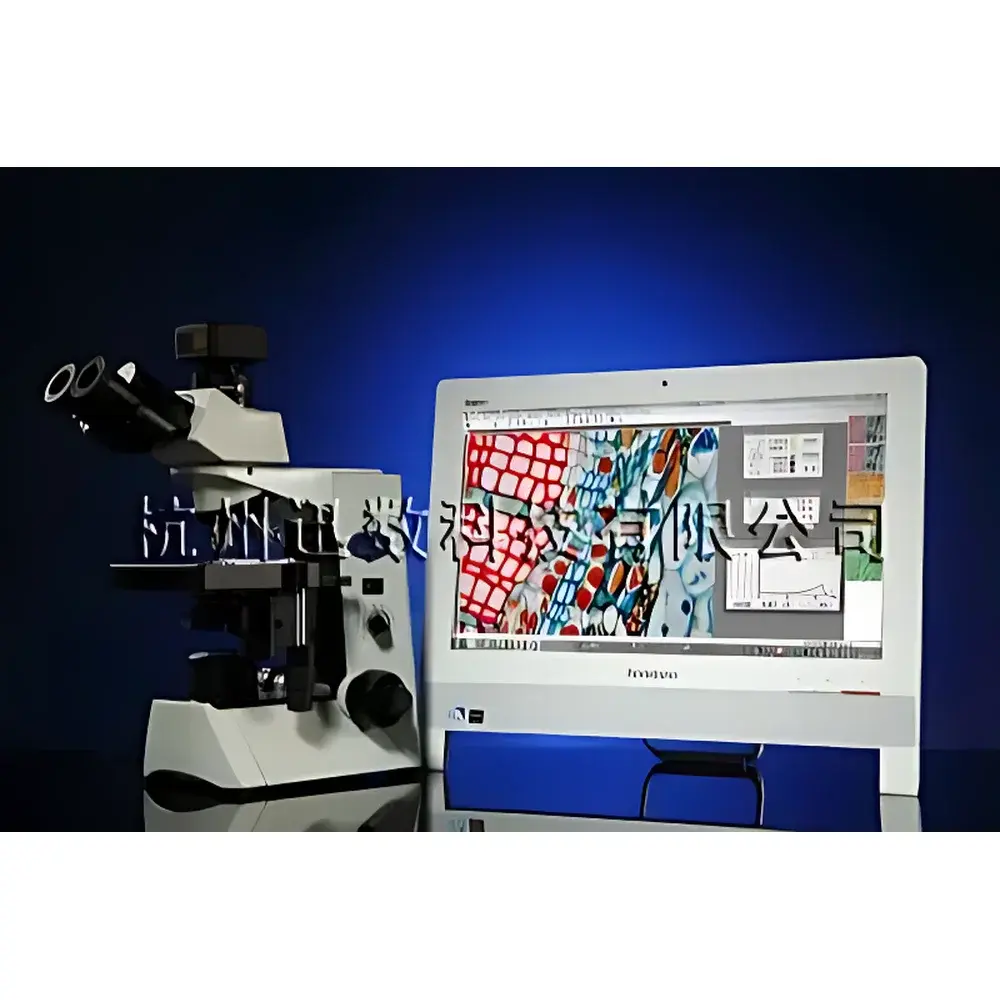
Shineso MIC Series Microscopic Image Analysis System
| Brand | Shineso |
|---|---|
| Origin | Zhejiang, China |
| Manufacturer Type | Original Equipment Manufacturer (OEM) |
| Country of Origin | China |
| Model | MIC Series (MIC-1 / MIC-2 / MIC-3) |
| Price Range | USD 2,800 – 7,000 (FOB) |
| Instrument Type | Upright Biological Microscope System |
| Optical Platform | Infinity-Corrected UIS Optical System (MIC-3) |
| Camera Resolution | 1280×1024 (MIC-1), 2048×1536 (MIC-2 & MIC-3) |
| Software | SHINESO-MIC Image Analysis Suite v5.x |
| Compliance | CE-marked for laboratory use |
Overview
The Shineso MIC Series Microscopic Image Analysis System is an integrated upright biological microscope platform engineered for quantitative digital microscopy in life science research, materials characterization, and industrial quality control. It combines a precision optical microscope—configured with infinity-corrected optics (UIS system in MIC-3 variant)—with a high-sensitivity color CMOS imaging sensor and a purpose-built image analysis software suite. The system operates on the principle of digital optical microscopy: light transmitted or reflected from the specimen is resolved by plan-apochromatic or semi-apochromatic objectives, captured by a calibrated CMOS sensor, and processed using deterministic algorithms for morphometric quantification. Unlike conventional eyepiece-based observation, the MIC system enables simultaneous real-time dynamic preview and static high-fidelity image acquisition—a capability enabled by Shineso’s proprietary Dual-Path Parallel Imaging Architecture. This architecture decouples live focusing feedback from analytical image capture, ensuring optimal focus stability during measurement and eliminating parallax-induced calibration drift common in single-path systems.
Key Features
- Triple-tier hardware configuration (MIC-1, MIC-2, MIC-3) supporting progressive performance scaling—from entry-level domestic trinocular microscopes to research-grade Olympus CX31 platforms with UIS infinity optics
- High-fidelity CMOS imaging sensors: 1.3 MP (1280×1024) resolution in MIC-1; 3.1 MP (2048×1536) in MIC-2/MIC-3, delivering spatial resolution down to 1.0–2.0 µm under optimal Köhler illumination
- Dual-path parallel imaging engine: Enables concurrent live video feed (for rapid focusing and stage navigation) and high-bit-depth static frame capture (for measurement-grade analysis) without temporal or geometric coupling
- Comprehensive image enhancement toolkit: Includes adaptive contrast enhancement, multi-mode background flattening, six configurable spatial filters (Gaussian, median, low-pass, etc.), and morphology-based edge-preserving sharpening
- Calibration-integrated metrology: On-system pixel-to-micron calibration with traceable reference standards; supports user-defined objective magnification presets and stage coordinate mapping
- Modular software architecture: All core functions—including image acquisition, preprocessing, segmentation, morphometry, statistical reporting, and database archiving—are implemented as independent, version-controlled modules compliant with ISO/IEC 17025 documentation requirements
Sample Compatibility & Compliance
The MIC Series accommodates standard 24×50 mm and 26×76 mm glass slides, coverslips (No. 1.5, 0.17 mm thickness), and petri dishes (up to 100 mm diameter) via adjustable mechanical stage with vernier scales. It supports brightfield, phase contrast, and basic polarized light observation modes—compatible with routine histological sections, stained cytology smears, microbial colonies, metallurgical grain structures, airborne particulates, and polymer phase domains. While not designed for fluorescence or confocal applications, its optical path preserves >92% transmission across 400–700 nm, enabling post-acquisition pseudo-color spectral analysis. The system meets CE safety directives (2014/30/EU EMC, 2014/35/EU LVD) and is validated for use in ISO 13485-certified medical device manufacturing environments. Optional audit-trail logging complies with FDA 21 CFR Part 11 requirements for electronic records and signatures when deployed in GxP workflows.
Software & Data Management
The SHINESO-MIC Image Analysis Suite (v5.x) is a Windows-native application built on a modular, event-driven framework. It features a non-destructive editing pipeline: all processing steps are logged as reversible operations with parameter metadata embedded in TIFF headers. The software includes full DICOM-SR export capability for integration into PACS environments and supports CSV/Excel batch export of morphometric tables (area, perimeter, Feret diameter, circularity, aspect ratio, RGB intensity histograms). A relational SQLite database stores annotated images, calibration profiles, user permissions, and session logs—with encrypted backups and role-based access control (RBAC) for multi-user labs. Report generation supports customizable templates compliant with ISO 10993 biocompatibility testing documentation and ASTM E2917-22 guidelines for particle size distribution reporting.
Applications
- Cell biology: Quantitative assessment of cell confluence, viability staining (e.g., Trypan Blue exclusion), mitotic index scoring, and neurite outgrowth measurement in primary neuronal cultures
- Hematology & microbiology: Automated blood smear differential counts, bacterial colony enumeration on agar plates, yeast/bacterial morphology classification using color-thresholded segmentation
- Materials science: Grain size distribution analysis per ASTM E112, inclusion counting in steel per ISO 4967, polymer crystallinity domain quantification
- Environmental monitoring: Airborne particulate matter (PM10/PM2.5) sizing and shape factor analysis from filter-captured samples
- Pharmaceutical QC: Excipient particle size uniformity testing in tablet granulations (per USP ), foreign particulate identification in parenteral solutions
FAQ
Does the MIC system support fluorescence imaging?
No—the optical train is optimized for transmitted brightfield and phase contrast. Fluorescence requires dedicated excitation/emission filters, dichroic mirrors, and cooled EMCCD/sCMOS sensors not included in this configuration.
Can measurement data be exported to LIMS or ELN platforms?
Yes—CSV, XML, and structured PDF exports are supported. API-based integration with LabVantage, Benchling, and IDBS E-WorkBook is available through optional middleware licensing.
Is the software validated for regulated environments?
The base software is IQ/OQ-qualified. Full 21 CFR Part 11 compliance—including electronic signature enforcement, change control, and audit trail review—requires activation of the Validation Support Package and documented site-specific PQ protocols.
What is the maximum field-of-view resolution achievable at 40× objective?
At 40× with the MIC-3’s Olympus UIS objective and 2048×1536 sensor, the effective pixel scale is 0.22 µm/pixel over a 450 µm × 340 µm FOV—enabling subcellular feature resolution consistent with ISO 10993-12 guidance.
How does the system handle overlapping or clustered particles?
The software implements watershed-based separation with adjustable seed point density and manual correction overlay. Users may apply intensity gradient thresholding, distance transform masking, or trainable Weka segmentation plugins (via Fiji bridge) for complex agglomerates.


