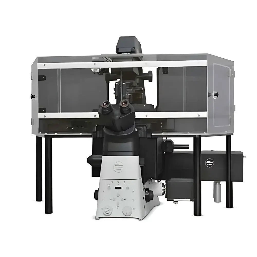
Nikon N-SIMS Super-Resolution Laser Confocal Microscope
| Brand | Nikon |
|---|---|
| Origin | Japan |
| Model | N-SIMS |
| Lateral Resolution (XY, FWHM) | 86 nm (TIRF-SIM), 115 nm (3D-SIM) |
| Axial Resolution (Z, FWHM) | 269 nm (3D-SIM) |
| Max Imaging Speed | 15 fps (TIRF-SIM/2D-SIM, 2 ms exposure) |
| Reconstruction Image Size | 1024 × 1024 or 2048 × 2048 pixels |
| Imaging Modes | TIRF-SIM, 2D-SIM, 3D-SIM (single-frame & time-series reconstruction) |
| Multicolor Capability | Up to 6 channels, simultaneous dual-color acquisition |
| Compatible Laser Units | LU-NV (std: 405/488/561/640 nm |
| opt | 445 nm), LUD-HV (std: same |
| opt | 445/515 nm), Ziva Light Engine (405/446/476/518/545/637 nm) |
| Compatible Microscope Platform | Nikon ECLIPSE Ti2-E motorized inverted microscope with Perfect Focus System (PFS), encoded motorized XY stage, and piezo-driven Z-stage |
| Objective Lenses | CFI SR HP Plan Apochromat Lambda S 100XC Sil (NA 1.35), CFI SR HP Apochromat TIRF 100XC Oil (NA 1.49), CFI SR HP Apochromat TIRF 100XAC Oil (NA 1.49), CFI SR Plan Apochromat IR 60XC WI (NA 1.27), CFI SR Plan Apochromat IR 60XAC WI (NA 1.27), CFI Plan Apochromat Lambda 60XC (NA 0.95), CFI Plan Apochromat Lambda 40XC (NA 0.95) |
| Camera | ORCA-Fusion BT sCMOS (Hamamatsu Photonics K.K.) |
| Software | NIS-Elements AR (with NIS-A 6D and N-SIM analysis modules required), NIS-Elements C (for AX/AX R confocal systems) |
| Operating Environment | 20–28 °C (±1.5 °C) |
Overview
The Nikon N-SIMS Super-Resolution Laser Confocal Microscope is a high-precision structured illumination microscopy (SIM) platform engineered for quantitative, multi-dimensional biological imaging beyond the diffraction limit. Leveraging optical patterned illumination and computational reconstruction, the N-SIMS implements three distinct SIM modalities—TIRF-SIM, 2D-SIM, and 3D-SIM—to achieve isotropic and anisotropic resolution enhancement in live and fixed specimens. Its core optical architecture is integrated into the Nikon ECLIPSE Ti2-E inverted microscope platform, featuring active drift compensation via the Perfect Focus System (PFS), sub-nanometer Z-axis control using a closed-loop piezo stage, and high-fidelity multichannel excitation compatibility across visible and near-UV spectral bands. Designed for rigorous cell biology, neuroimaging, and structural proteomics workflows, the N-SIMS delivers reproducible sub-100 nm lateral resolution under TIRF-SIM conditions and maintains robust axial discrimination in thick samples through optimized 3D-SIM reconstruction algorithms.
Key Features
- Sub-diffraction resolution: 86 nm lateral (XY) resolution in TIRF-SIM mode and 115 nm in 3D-SIM mode, validated using fluorescent microspheres (FWHM measurement); 269 nm axial (Z) resolution in 3D-SIM mode
- High-speed acquisition: Up to 15 frames per second in TIRF-SIM and 2D-SIM configurations using 2 ms exposure times, enabling dynamic visualization of intracellular trafficking and cytoskeletal remodeling
- Flexible multicolor imaging: Support for up to six fluorescence channels with simultaneous dual-color acquisition, compatible with Nikon’s LU-NV, LUD-HV, and Ziva Light Engine laser units spanning 405–637 nm
- Advanced hardware integration: Motorized Ti2-E platform with encoded XY stage, PFS-based focus stabilization, and piezo Z-stage for precise optical sectioning and long-term time-lapse stability
- Optimized objective suite: Includes CFI SR HP Apochromat TIRF 100XC Oil (NA 1.49) for evanescent-field imaging and CFI SR HP Plan Apochromat Lambda S 100XC Sil (NA 1.35) for deep-tissue SIM with reduced spherical aberration
- High-sensitivity detection: Hamamatsu ORCA-Fusion BT sCMOS camera delivering low-noise, large-field-of-view imaging at 1024 × 1024 or 2048 × 2048 pixel reconstruction formats
Sample Compatibility & Compliance
The N-SIMS supports a broad range of biological specimens—including adherent and suspension mammalian cells, primary neurons, organoids, and thin tissue sections—without requiring specialized labeling chemistry beyond conventional fluorescent probes (e.g., Alexa Fluor, ATTO, and SiR dyes). Its TIRF-SIM configuration is optimized for plasma membrane and cortical cytoskeleton studies, while 3D-SIM enables volumetric nanoscale mapping in specimens up to ~15 µm thick when combined with refractive-index-matched mounting media. The system complies with ISO 19012-1:2021 (microscopy—nomenclature and definitions) and supports GLP/GMP-aligned documentation protocols when paired with NIS-Elements AR’s audit-trail-enabled software modules. All optical components meet JIS B 7151 standards for precision optical alignment and thermal stability.
Software & Data Management
Acquisition, reconstruction, and quantitative analysis are managed through Nikon’s NIS-Elements AR platform, extended with mandatory NIS-A 6D and N-SIM analysis modules. These modules provide real-time SIM reconstruction, channel registration, deconvolution-assisted artifact suppression, and batch processing for time-series and multivolume datasets. NIS-Elements AR supports FDA 21 CFR Part 11-compliant user authentication, electronic signatures, and immutable audit trails when deployed in regulated environments. Raw data are stored in TIFF or ND2 format with embedded metadata (wavelength, exposure, stage position, objective ID), ensuring traceability and FAIR (Findable, Accessible, Interoperable, Reusable) data principles. Export options include standardized OME-TIFF and HDF5 containers for interoperability with Python-based analysis pipelines (e.g., scikit-image, napari).
Applications
- Nanoscale organelle architecture: Quantitative mapping of mitochondrial cristae, nuclear pore complexes, and endosomal sorting machinery
- Single-molecule localization correlation: Combined use with PALM/STORM for correlative super-resolution workflows
- Live-cell dynamics: High-temporal-resolution tracking of clathrin-coated pit assembly, actin polymerization fronts, and synaptic vesicle recycling
- Multiplexed protein co-localization: Simultaneous 4–6 color imaging of post-translational modification gradients (e.g., phospho-ERK, ubiquitin, SUMO)
- 3D ultrastructural phenotyping: Automated segmentation and morphometric analysis of microtubule networks in mitotic spindles or neuronal dendritic arbors
FAQ
What is the minimum required sample preparation for optimal N-SIMS performance?
Standard immunofluorescence or live-cell dye labeling suffices; however, optimal results require high signal-to-noise ratio, minimal out-of-focus background, and refractive index matching for 3D-SIM acquisitions.
Is the N-SIMS compatible with expansion microscopy (ExM) protocols?
Yes—the system supports ExM-treated samples imaged with standard objectives, though immersion-matched objectives (e.g., CFI SR HP Apochromat TIRF 100XAC Oil) are recommended for maximal resolution recovery.
Can N-SIMS data be exported for third-party analysis in MATLAB or Python?
Yes—ND2 and OME-TIFF exports retain full metadata and spatial calibration, enabling direct import into open-source toolkits such as NanoJ, SIMcheck, or custom Fourier-domain reconstruction scripts.
Does the system support automated Z-stack acquisition during SIM reconstruction?
Yes—time-lapse 3D-SIM sequences can be programmed with synchronized Z-stepping, piezo positioning, and multi-channel illumination timing via NIS-Elements AR scripting interface.
What environmental controls are recommended for stable long-term operation?
A temperature-stabilized room (20–28 °C ±1.5 °C) with vibration isolation and low-EMI power conditioning is required; optional active air-table integration is supported for sub-50 nm stability in extended acquisitions.


