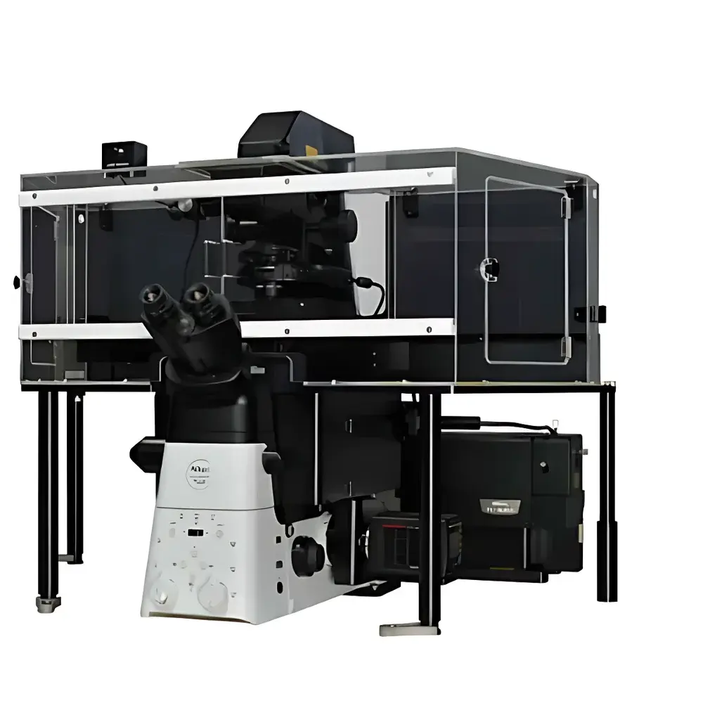
Nikon N-SIM E Super-Resolution Microscope
| Brand | Nikon |
|---|---|
| Origin | Japan |
| Model | N-SIM E |
| Resolution (XY) | ~115 nm |
| Resolution (Z, 3D-SIM) | ~300 nm |
| Imaging Speed | ~1 frame/sec |
| Field of View | 66 µm × 66 µm |
| Objective Compatibility | High-NA objectives up to NA 1.49 |
| Modality | Structured Illumination Microscopy (SIM) with integrated confocal capability |
| 3D Reconstruction Modes | Slice and Stack |
| Compliance | Designed for GLP/GMP-adjacent research environments |
Overview
The Nikon N-SIM E is a streamlined, entry-tier super-resolution microscope engineered for laboratories seeking accessible, high-fidelity structural imaging beyond the diffraction limit—without the operational complexity or cost premium associated with single-molecule localization microscopy (SMLM) or stimulated emission depletion (STED) platforms. It implements widefield-based structured illumination microscopy (SIM), a deterministic, linear optical technique that computationally extracts high-spatial-frequency information by acquiring multiple phase-shifted, patterned illumination images. Unlike stochastic methods requiring sparse fluorophore activation, SIM operates under conventional fluorescence labeling protocols and standard laser power levels, making it intrinsically suitable for live-cell applications where phototoxicity and photobleaching must be minimized. The system integrates natively with Nikon’s Eclipse Ti2 inverted microscope platform and leverages proprietary high-numerical-aperture (NA ≤ 1.49) CFI Apo TIRF objectives to achieve a lateral resolution of approximately 115 nm—nearly double that of conventional widefield epifluorescence—and axial resolution down to ~300 nm in 3D-SIM mode. Its design prioritizes reproducibility, ease of alignment, and compatibility with routine cell culture workflows.
Key Features
- Diffraction-unlimited imaging at ~115 nm lateral resolution using SIM principles—no specialized dyes or buffer systems required
- Real-time acquisition capability: up to 1 frame per second in 2D-SIM mode, enabling dynamic observation of subcellular processes including vesicle trafficking, cytoskeletal remodeling, and organelle interactions
- 66 µm × 66 µm field of view in super-resolution mode—significantly larger than typical STED or PALM/STORM fields—supporting high-throughput screening of neuronal networks, tissue sections, or multi-cellular assays
- Dual 3D-SIM reconstruction algorithms: “Slice” mode delivers optically sectioned images with enhanced axial contrast for thin specimens (e.g., adherent monolayers); “Stack” mode applies Gustafsson-derived deconvolution for thicker samples (up to ~10 µm), preserving signal-to-noise ratio across depth
- Seamless modality switching: users may acquire confocal reference images via integrated laser scanning unit, then instantly reposition and acquire SIM data from the same region—enabling direct correlation between diffraction-limited context and super-resolved detail
- Automated calibration and drift compensation: hardware-synchronized stage and focus control minimize positional artifacts during time-lapse SIM acquisitions
Sample Compatibility & Compliance
The N-SIM E supports standard fluorescent probes—including GFP, mCherry, Alexa Fluor dyes, and SiR-tubulin—across fixed and live mammalian, insect, and plant specimens. It accommodates common mounting media (e.g., ProLong Diamond, Vectashield) and glass-bottom dishes (No. 1.5 coverslip thickness). The system complies with ISO 10993 biocompatibility guidelines for optical components in contact with biological samples and meets CE marking requirements for laboratory instrumentation. While not a regulated medical device, its architecture supports audit-ready operation in GLP- and GMP-aligned research settings: raw image metadata (including illumination pattern parameters, exposure timing, objective ID, and environmental logs) are embedded in TIFF headers and preserved through NIS-Elements AR export pipelines. When deployed with validated software configurations and electronic signature modules, the workflow satisfies traceability criteria outlined in FDA 21 CFR Part 11 for non-clinical study data.
Software & Data Management
NIS-Elements AR (Advanced Research) v5.0+ serves as the unified acquisition and processing engine. It provides real-time SIM reconstruction, batch processing of time-series datasets, and export of calibrated, metadata-rich TIFF stacks compliant with OME-TIFF standards. Quantitative analysis modules include colocalization (Pearson’s r, Manders’ coefficients), intensity profiling, and object-based segmentation—validated against reference datasets from the BioImage Archive. All processing steps are logged with timestamps, user IDs, and parameter sets, enabling full experimental provenance. Data exports support integration with third-party platforms such as Fiji/ImageJ (via open-source SIM plugins), Imaris (for surface rendering), and MATLAB for custom algorithm development. Raw patterned image sequences are retained alongside reconstructed outputs, ensuring full reproducibility and reprocessing flexibility.
Applications
- Subcellular architecture mapping: visualization of nuclear pore complexes, endoplasmic reticulum tubules, mitochondrial cristae, and synaptic protein clusters
- Live-cell dynamics: tracking of clathrin-coated pit formation, microtubule plus-end dynamics, and chromatin condensation during mitosis
- Neuroscience: high-content imaging of dendritic spines, axonal boutons, and co-distribution of pre- and postsynaptic markers across extended neuronal fields
- Cell migration studies: quantification of focal adhesion turnover and actin flow patterns in wound-healing assays
- Pathogen-host interactions: spatial resolution of viral replication factories or bacterial inclusion bodies within infected cells
FAQ
What is the minimum recommended fluorophore brightness for reliable N-SIM E imaging?
Fluorophores should exhibit ≥10⁶ photons per molecule per second under 488 nm excitation at ≤10 mW/mm² irradiance. GFP-tagged constructs expressed at endogenous or moderate overexpression levels typically meet this threshold.
Can N-SIM E be upgraded to support TIRF-SIM or multi-color SIM?
Yes—the system’s modular optical path supports optional TIRF illuminators and multi-line laser combiners. Multi-color SIM requires sequential acquisition with spectral unmixing; no hardware modification is needed beyond appropriate filter sets and dichroics.
Is drift correction available during long-term 3D-SIM time-lapse?
Yes—hardware-based Z-drift compensation (using Nikon’s Perfect Focus System v4.0) and XY stage stabilization (via motorized piezo stage feedback) are enabled by default in time-lapse SIM acquisition protocols.
How does N-SIM E handle out-of-focus light compared to confocal?
Unlike point-scanning confocal, N-SIM E uses computational optical sectioning via patterned illumination and Fourier-domain reconstruction. While not rejecting out-of-focus light physically, it achieves comparable axial discrimination (≈300 nm) through frequency-domain filtering—retaining higher photon efficiency than confocal for equivalent SNR.
What file formats are supported for raw data export and archival?
Raw pattern images and reconstructed stacks are saved as multi-page TIFFs with embedded OME-XML metadata. NIS-Elements also supports HDF5 export for large-scale dataset storage and HPC pipeline ingestion.


