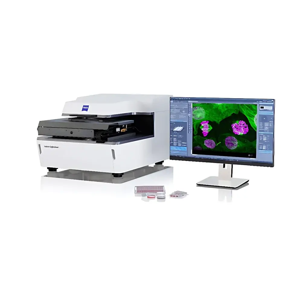
ZEISS Lattice Lightsheet 7 Live-Cell Light-Sheet Microscopy System
| Brand | ZEISS |
|---|---|
| Origin | Germany |
| Model | Lattice Lightsheet 7 |
| Import Status | Imported Instrument |
| Distribution Type | Authorized Distributor |
Overview
The ZEISS Lattice Lightsheet 7 is a high-performance, fully integrated light-sheet fluorescence microscope engineered for long-term, low-phototoxicity, high-resolution 3D imaging of living biological specimens. Unlike conventional Gaussian light-sheet systems, it employs lattice light-sheet illumination—a technique based on structured illumination using a 2D optical lattice generated via spatial light modulation and adaptive optics. This produces an ultrathin, quasi-diffraction-limited light sheet (typically < 0.5 µm thick at the focal plane), enabling near-isotropic spatial resolution (~250–300 nm lateral, ~350–450 nm axial) while maintaining high signal-to-noise ratio and minimal out-of-focus excitation. The system operates on the principle of orthogonal illumination and detection: the excitation light sheet propagates perpendicular to the detection axis, eliminating axial photobleaching and enabling volumetric acquisition at speeds up to 100–300 volumes per second—depending on field-of-view and pixel binning—without compromising viability. Designed for sustained live-cell observation, it supports time-lapse imaging over hours to days, making it ideal for developmental biology, organoid dynamics, mitotic progression, and subcellular trafficking studies under physiologically relevant conditions.
Key Features
- Ultrathin lattice light-sheet generation via real-time adaptive beam shaping, delivering sub-500 nm effective sheet thickness and reduced scattering in optically heterogeneous samples
- Integrated dual-side illumination path with automatic beam alignment and aberration correction for uniform illumination across large fields of view (up to 1.2 mm × 1.2 mm)
- Motorized, multi-axis sample positioning stage with nanometer-level repeatability and closed-loop feedback control for precise multi-position time-lapse experiments
- Automated optical calibration routine executed prior to each session—compensating for thermal drift, refractive index mismatches, and immersion medium variations
- Modular optical design supporting multiple excitation lasers (405, 488, 561, 640 nm standard; optional UV/IR extensions) and highly sensitive sCMOS or EMCCD detection pathways
- Native compatibility with standard culture vessels: glass-bottom dishes, µ-slides, capillaries, and agarose-embedded specimens—no custom mounting required
Sample Compatibility & Compliance
The Lattice Lightsheet 7 accommodates a broad range of biological preparations without requiring specialized embedding or clearing protocols. It supports live single cells (e.g., LLC-PK1, HeLa, iPSCs), 3D cell cultures (organoids, spheroids, hydrogels), early-stage model organisms (zebrafish embryos, C. elegans, Drosophila melanogaster), and expanded tissue specimens (expansion microscopy samples). All optical paths are aligned to ISO 10110-3 standards for wavefront fidelity, and mechanical stability meets ISO 10360-2 for coordinate measuring systems. The system complies with IEC 61000-6-3 (EMC emission) and IEC 61000-6-2 (immunity) standards. For regulated environments, audit trails, user access controls, and electronic signatures can be enabled in accordance with FDA 21 CFR Part 11 requirements when paired with ZEISS ZEN Blue v3.6+ software in GLP/GMP mode.
Software & Data Management
Acquisition, processing, and analysis are unified within ZEISS ZEN Blue (v3.6 or later), a modular platform supporting automated experiment scripting, real-time deconvolution, and GPU-accelerated rendering. Raw data are stored in open, metadata-rich formats (OME-TIFF, HDF5) compliant with the OME data model and FAIR principles. Built-in tools include multichannel registration, intensity normalization across time points, 4D segmentation (via AI-assisted trainable Weka classifiers), and quantitative colocalization analysis (Manders’ coefficients, Pearson correlation). Data export supports interoperability with Imaris, Fiji/ImageJ, MATLAB, and Python-based analysis pipelines (e.g., napari, scikit-image). Optional ZEISS Lab Archive integration enables centralized storage, version-controlled project management, and role-based access across multi-user facilities.
Applications
- Long-term live-cell imaging of nuclear lamina dynamics (e.g., LMNB1-mEGFP in human iPSCs), mitotic spindle assembly, and chromosome segregation
- High-speed volumetric tracking of organelle motility (mitochondria, lysosomes, ER) in intact zebrafish embryos or mouse oocytes arrested at metaphase II
- Structural and functional phenotyping of brain organoids—monitoring neuronal migration, synapse formation, and calcium wave propagation over days
- Quantitative assessment of drug-induced morphological changes in 3D tumor spheroids under controlled perfusion conditions
- Post-expansion microscopy (ExM) 3D reconstruction of synaptic ultrastructure with molecular specificity and isotropic resolution
FAQ
What types of immersion media are supported?
The system is optimized for aqueous-based media (e.g., embryo medium, Leibovitz’s L-15, PBS) and compatible with silicone oil (n = 1.40), water (n = 1.33), and glycerol (n = 1.47) immersion objectives. Custom refractive index matching is configurable via ZEN software.
Can the Lattice Lightsheet 7 be integrated into existing incubation setups?
Yes—it features standardized mounting interfaces and environmental control ports (for temperature, CO₂, and humidity monitoring) compatible with commercial stage-top incubators and chamber systems.
Is multi-color imaging supported with spectral unmixing?
Yes—ZEN Blue includes linear unmixing algorithms for up to five spectrally distinct fluorophores, leveraging reference spectra acquired during setup or imported from public databases (e.g., FPbase, Fluorophore.org).
What is the maximum achievable imaging depth in scattering tissues?
In cleared or lightly scattering samples (e.g., early zebrafish embryos, thin organoids), usable depth exceeds 150 µm. With adaptive optics correction and lattice illumination optimization, effective depth in moderately scattering specimens (e.g., PFA-fixed brain slices post-clearing) reaches ~80–100 µm at Nyquist-sampled resolution.
Does the system support light-sheet–based optogenetics?
Yes—the illumination path can be temporally gated and spatially patterned via the SLM, enabling targeted photoactivation or photoinhibition synchronized with image acquisition—fully programmable through ZEN scripting.


