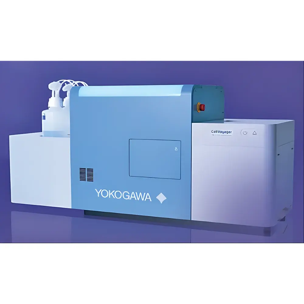
Yokogawa CellVoyager CQ3000 High-Content Analysis System
| Brand | Yokogawa |
|---|---|
| Origin | Japan |
| Model | CQ3000 |
| Configuration | Desktop-integrated HCA platform with CSU-based confocal imaging, dual-camera option, sCMOS detector, integrated environmental chamber (7-day live-cell incubation), uniform illumination module, high-NA water-immersion objective, target-search automation, and 100 fps high-speed acquisition mode |
| Compliance | Designed for GLP/GMP-aligned workflows |
| Software | CellPathfinder (AI-assisted image analysis with deep learning modules) |
Overview
The Yokogawa CellVoyager CQ3000 is a benchtop high-content analysis (HCA) system engineered for quantitative, multi-parametric phenotypic profiling of fixed and live cells under physiologically relevant conditions. At its core, the system integrates Yokogawa’s proprietary Confocal Scanner Unit (CSU) — a spinning-disk confocal architecture — enabling optical sectioning with minimal phototoxicity and photobleaching. This principle ensures high-fidelity 3D reconstruction of thick specimens such as organoids, tissue slices, and microphysiological systems (MPS), while maintaining cellular viability over extended observation periods (up to 7 days). The CQ3000 combines rapid volumetric imaging (up to 100 frames per second in high-speed mode), uniform Köhler-style illumination across the full field-of-view (FOV), and hardware-synchronized dual-channel acquisition via optional dual sCMOS cameras. Its compact footprint and modular design support deployment in core facilities, academic labs, and regulated biopharma environments where reproducibility, traceability, and scalability are critical.
Key Features
- Spinning-disk confocal imaging using Yokogawa CSU technology for low-phototoxicity, high-speed optical sectioning
- Integrated environmental chamber with precise CO2, temperature, and humidity control for long-term live-cell assays (≥168 hours)
- High numerical aperture (NA) water-immersion objective optimized for clear visualization of superficial layers in thick samples (e.g., brain slices, tumor spheroids, organoids)
- Dual-camera configuration supporting simultaneous two-color acquisition without spectral crosstalk or time-lag
- Uniform illumination module enabling consistent signal intensity across multi-field stitched acquisitions — essential for whole-tissue-section analysis
- Target-search automation: low-magnification survey scan followed by AI-guided high-magnification reimaging of detected objects (e.g., mitotic cells, fluorescently labeled organelles, or rare phenotypes)
- High-speed acquisition mode (up to 100 fps) suitable for dynamic processes including cardiomyocyte contraction, calcium transients, and vesicle trafficking
- sCMOS detector with high quantum efficiency (>80% at 550 nm) and low read noise (<1.5 e−) for superior signal-to-noise ratio in low-light conditions
Sample Compatibility & Compliance
The CQ3000 accommodates standard microplate formats (6–1536-well), glass-bottom dishes, chamber slides, and custom substrates used in 3D cell culture and organ-on-a-chip applications. It supports fixation protocols (paraformaldehyde, methanol), live-cell dyes (Calcein AM, Hoechst 33342, Fluo-4), immunofluorescence labeling, and Cell Painting multiplexed staining workflows. Hardware and software are designed to meet requirements for regulated research environments: metadata-rich OME-TIFF output ensures FAIR data principles; audit trails and user-access logs align with GLP documentation standards; API interfaces (RESTful) enable bidirectional communication with laboratory information management systems (LIMS) and electronic lab notebooks (ELN). While not certified as medical device hardware, the system adheres to ISO 13485-aligned manufacturing practices and supports 21 CFR Part 11-compliant software configurations when deployed with validated CellPathfinder analysis modules.
Software & Data Management
CellPathfinder is the dedicated analysis suite for the CQ3000, featuring GPU-accelerated segmentation, deep learning–based object classification (trained on user-supplied ground-truth datasets), and hierarchical morphometric quantification (e.g., nuclear texture, mitochondrial network fragmentation, neurite outgrowth metrics). All analyses generate structured CSV/JSON reports alongside interactive HTML dashboards. Raw and processed data are exported in open, community-standard formats: OME-TIFF for multi-dimensional image stacks and OMERO-compatible annotations for collaborative annotation and review. Batch processing pipelines can be scripted via Python SDK, and analysis workflows are version-controlled and exportable for cross-lab reproducibility. No proprietary binary lock-in is imposed — all intermediate files remain fully accessible and interpretable.
Applications
- Organoid and spheroid phenotyping: quantifying size distribution, lumen formation, and differentiation markers across 3D cultures
- Toxicology screening: real-time assessment of mitochondrial membrane potential, lysosomal pH, and DNA damage response kinetics
- Neuroscience: automated tracing of dendritic spines, synapse counting, and activity-dependent structural plasticity
- Cardiac safety pharmacology: beat-rate variability, calcium transient amplitude, and conduction velocity mapping in iPSC-derived cardiomyocytes
- Immunooncology: spatial analysis of immune cell infiltration, checkpoint protein co-expression, and tumor-stroma interactions in co-culture models
- Drug mechanism-of-action studies using Cell Painting: unsupervised clustering of morphological profiles to infer compound target pathways
FAQ
What types of microplates and sample carriers are supported?
Standard ANSI/SLAS-compliant plates (6–1536-well), glass-bottom 35 mm dishes, µ-Slide chambers (Ibidi), and custom-engineered organ-chips with transparent membranes.
Can the CQ3000 perform time-lapse imaging over multiple days without focus drift?
Yes — the system includes hardware autofocus (infrared-based) and thermal stabilization to maintain Z-position accuracy within ±0.3 µm over 168-hour acquisitions.
Is CellPathfinder compatible with third-party deep learning frameworks like TensorFlow or PyTorch?
CellPathfinder supports model import/export in ONNX format; users may train custom CNN/U-Net architectures externally and deploy them directly into the analysis pipeline.
How does the target-search function reduce data storage burden?
By limiting high-resolution imaging to only regions containing predefined morphological or intensity criteria — reducing total image volume by up to 92% compared to full-FOV acquisition.
Does the system support multi-user access with role-based permissions?
Yes — administrator-configurable user roles (operator, analyst, reviewer) govern instrument control, analysis parameter editing, and report approval workflows.


