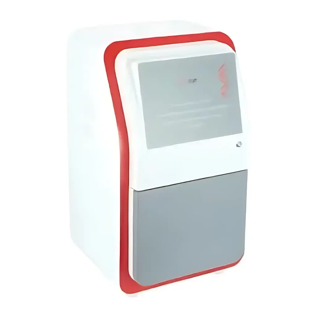
Jiapeng JP-K3900 Chemiluminescence Gel Imaging System
| Brand | Jiapeng |
|---|---|
| Origin | Shanghai, China |
| Model | JP-K3900 |
| Imaging Modality | Chemiluminescence, Fluorescence (UV/Blue/Green/Red), White Light Transmission & Reflection |
| CCD Sensor | Sony IMX533CLK-D, 4/3-inch format, 3000 × 3000 (9 MP) |
| Pixel Size | 5.4 µm × 5.4 µm |
| Bit Depth | 16-bit (0–65,535 grayscale) |
| Dynamic Range | >4.8 OD |
| Cooling Temperature | −45 °C (ΔT ≤ −65 °C vs ambient) |
| Quantum Efficiency | >75% |
| Lens | Computar F/0.95, 8–48 mm motorized zoom lens |
| Signal-to-Noise Ratio | ≥56 dB |
| Detection Sensitivity | Protein ≥0.02 ng, DNA ≥0.05 ng (chemiluminescent substrates) |
| Dark Current | <0.0005 e⁻/pixel/s |
| Transmission UV Sources | 302 nm (standard), 254 nm & 365 nm (optional reflectance) |
| UV Transilluminator | 200 × 200 mm, high-intensity |
| White Light Source | Top-mounted, uniform LED panel with >90% homogeneity |
| Sample Stage | Dual-position drawer-style platform (9.5 × 9.5 cm and 12.5 × 12.5 cm imaging areas) |
| Filter Wheel | Motorized 5-position (standard), optional 10-position |
| Power Supply | AC 220 V, 50/60 Hz |
| Dimensions (W × D × H) | 390 × 440 × 735 mm |
| Weight | 18.3 kg |
Overview
The Jiapeng JP-K3900 Chemiluminescence Gel Imaging System is a fully automated, multi-modal digital imaging platform engineered for quantitative analysis of nucleic acids and proteins across electrophoretic gels, blots, and microplates. It operates on the principle of photon capture via a thermoelectrically cooled scientific CMOS sensor, optimized for low-light detection in chemiluminescent, fluorescent (UV, blue, green, red), and white-light transmission/reflection modes. Unlike conventional CCD-based systems, the JP-K3900 integrates a Sony IMX533CLK-D back-illuminated sensor with deep-cooling capability (−45 °C operational, up to −65 °C differential), enabling extended exposure times without thermal noise accumulation—critical for detecting weak chemiluminescent signals from low-abundance targets such as phosphorylated kinases or rare transcription factors. Its optical architecture supports both contact imaging (e.g., membrane-based Western blots) and non-contact fluorescence (e.g., SYBR Safe-stained gels), conforming to standard laboratory workflows in molecular biology, immunology, and functional genomics.
Key Features
- High-sensitivity cooled imaging: Sony IMX533CLK-D sensor with 4/3-inch format, 9 MP resolution (3000 × 3000), and quantum efficiency >75% at 550 nm—optimized for chemiluminescent signal capture.
- F/0.95 motorized zoom lens (Computar 8–48 mm) ensures maximal photon throughput and consistent focus across variable sample sizes; optional F/0.8 lens available for ultra-low-light applications.
- Dual-area drawer-style sample stage (9.5 × 9.5 cm and 12.5 × 12.5 cm) enables rapid switching between small-format membranes and large gels without manual repositioning.
- Integrated top-mounted white LED illumination with >90% intensity uniformity across field-of-view—ideal for documentation of Coomassie- or Ponceau S-stained membranes under visible light.
- Motorized 5-position filter wheel (standard), programmable via software; supports automatic filter selection synchronized with excitation source activation.
- Programmable auto-shutdown for UV and white-light sources (0–60 min timer), reducing photobleaching and operator exposure risk.
- Sealed dark chamber with anti-scatter interior coating minimizes stray light interference—essential for maintaining contrast in low-signal chemiluminescence acquisitions.
Sample Compatibility & Compliance
The JP-K3900 accommodates standard electrophoretic formats including mini and midi gels (up to 12.5 × 12.5 cm), nitrocellulose and PVDF membranes, microtiter plates (96-well), and stained tissue sections. Its UV transilluminator (200 × 200 mm, 302 nm) complies with IEC 61000-4-3 for electromagnetic compatibility and meets ISO 15195:2019 requirements for analytical instrument validation in clinical laboratories. While not FDA 510(k)-cleared, the system supports GLP-compliant documentation when paired with audit-trail-enabled acquisition software—enabling traceability of exposure time, gain, binning, filter selection, and metadata tagging per image file. All firmware and control logic adhere to IEC 62304 Class B software safety standards.
Software & Data Management
Bundled acquisition and analysis software provides full control over hardware parameters—including exposure duration (1 ms to 60 min), analog gain (1×–4×), pixel binning (1×1 to 8×8), and real-time background subtraction. Image files are saved in TIFF 16-bit format with embedded EXIF metadata (timestamp, lens focal length, temperature, filter ID). Quantitative modules support lane profiling, band intensity normalization against internal controls, molecular weight estimation using calibration ladders, and comparative densitometry across multiple exposures. Export options include CSV for statistical packages (R, GraphPad Prism), PDF reports with embedded LUTs, and DICOM-SR for integration into institutional PACS environments where permitted. Software versioning and update logs are retained for regulatory review.
Applications
- Western blot quantification using HRP- or AP-conjugated secondary antibodies with luminol- or CDP-Star-based substrates.
- DNA/RNA gel documentation under UV (302 nm) and ethidium bromide, SYBR Gold, or GelRed staining.
- Fluorescent protein detection (e.g., GFP, RFP, Cy5) on SDS-PAGE gels or membranes with appropriate excitation/emission filtering.
- Chemiluminescent ELISA and reporter gene assays (luciferase, β-galactosidase) in 96-well plate format.
- Stain-free total protein normalization via trihalo compound-based UV activation prior to immunodetection.
- Long-exposure bioluminescence imaging of live-cell reporters in low-oxygen environments (e.g., hypoxia-inducible factor studies).
FAQ
What is the minimum detectable amount of protein using chemiluminescence on the JP-K3900?
The system achieves a detection limit of ≥0.02 ng of bovine serum albumin (BSA) labeled with horseradish peroxidase (HRP) and developed with enhanced chemiluminescent substrate under standard acquisition settings (5-min exposure, F/0.95 lens, −45 °C sensor temperature).
Can the JP-K3900 perform multiplex fluorescent imaging?
Yes—the motorized filter wheel and programmable excitation source sequencing enable sequential acquisition of up to four fluorophores (e.g., DAPI, FITC, TRITC, Cy5) with spectral unmixing supported in post-processing.
Is the software compliant with 21 CFR Part 11 requirements?
The base software does not include electronic signature or audit-trail functionality out-of-the-box; however, an optional validated software package with role-based access control, electronic signatures, and immutable audit logs is available for GxP-regulated environments.
What maintenance is required for long-term calibration stability?
Annual verification of UV intensity output (using NIST-traceable radiometer), lens focus calibration, and dark-frame acquisition at −45 °C are recommended. No routine CCD replacement is necessary—the IMX533 sensor has a rated lifetime exceeding 10 years under typical lab use.
Does the system support third-party analysis plugins?
Yes—TIFF export with standardized metadata allows direct import into ImageJ/Fiji, MATLAB, and Python-based analysis pipelines (e.g., scikit-image, OpenCV) without proprietary format conversion.


