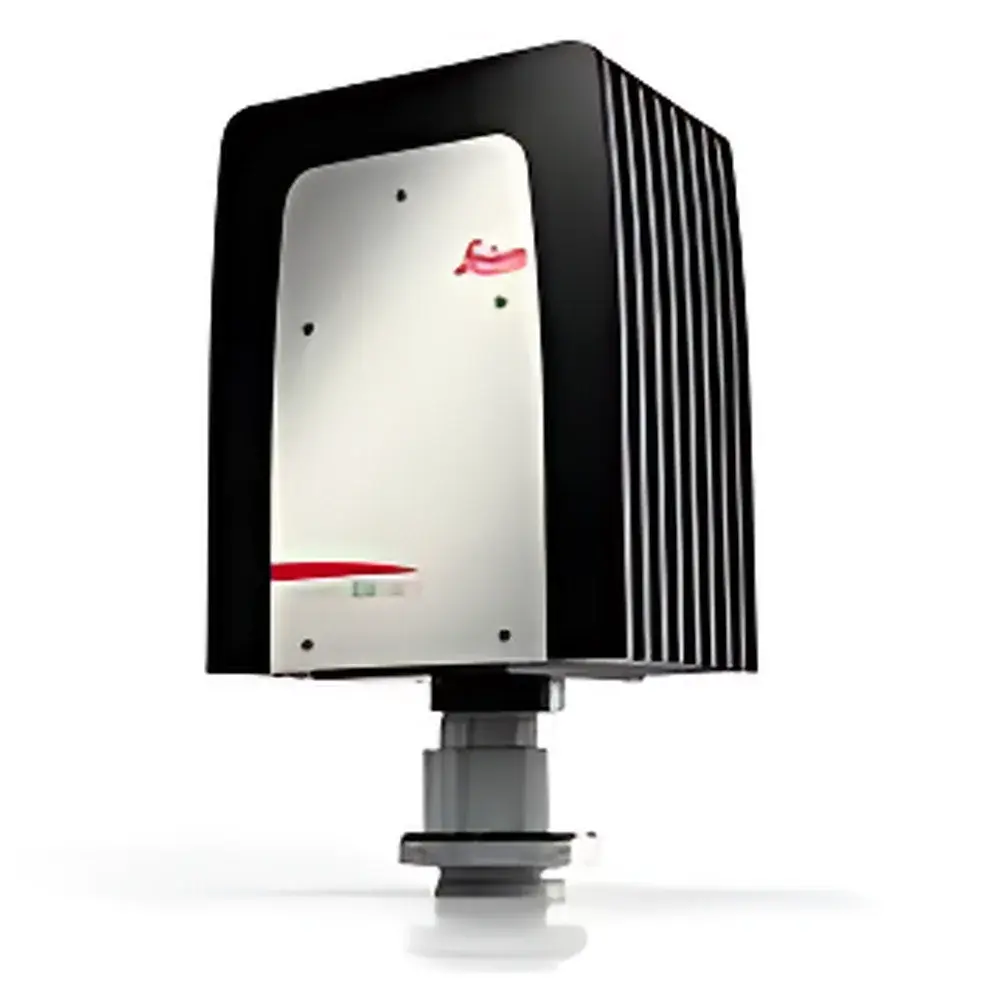
Leica DFC7000 T Digital Microscope Camera
| Brand | Leica |
|---|---|
| Origin | Germany |
| Model | DFC7000 T |
| Sensor Type | Progressive-scan CCD |
| Resolution | 2.8 MP (1920 × 1440) |
| Pixel Size | 4.54 µm |
| Cooling | Thermoelectric (Peltier), fanless operation |
| Dynamic Range | >64 dB (typ.) |
| Frame Rate | Up to 40 fps at full resolution |
| Color Interpolation | Adaptive 5×5 demosaicing with illumination-aware color correction |
| Monochrome Mode | Per-pixel luminance readout |
| Multi-channel Support | Simultaneous RGB channel acquisition |
| Operating Temperature Range | +5°C to +50°C |
| Interface | USB 3.0 |
| Compliance | CE, RoHS, FDA 21 CFR Part 11–ready software integration |
Overview
The Leica DFC7000 T is a high-performance, thermoelectrically cooled digital microscope camera engineered for demanding biological imaging applications across brightfield, fluorescence, and live-cell microscopy. Built around a progressive-scan CCD sensor with 2.8 megapixels (1920 × 1440) and 4.54 µm pixels, it delivers exceptional spatial resolution and quantum efficiency—particularly critical in low-light fluorescence modalities including DAPI, FITC, TRITC, Cy3, Cy5, and Cy5.5. Its optical design enables wide-field imaging without compromise, leveraging both native sensor geometry and compatibility with Leica’s low-magnification, high-NA objective lenses. Unlike conventional interline or rolling-shutter CMOS cameras, the DFC7000 T employs global shutter acquisition and correlated double sampling (CDS) to suppress read noise and fixed-pattern artifacts—ensuring quantitative fidelity in time-lapse, kinetic, and multi-channel co-localization experiments.
Key Features
- Thermoelectric cooling (Peltier-based) with passive heat dissipation—enabling fanless, vibration-free operation ideal for sensitive live-cell and long-exposure acquisitions.
- Adaptive 5×5 demosaicing algorithm that dynamically adjusts color interpolation based on illumination spectrum and intensity—preserving biologically accurate hue representation in brightfield histology and immunostained tissue sections.
- Dual-mode acquisition: seamless switching between full-color RGB mode and monochrome luminance mode, with independent pixel-level red, green, and blue channel readout for spectral unmixing and ratiometric analysis.
- Real-time frame rates up to 40 fps at full resolution (1920 × 1440), and up to 120 fps in binned 5×5 subregion mode—supporting rapid focus optimization, dynamic organelle tracking, and high-throughput screening workflows.
- Extended operating temperature range (+5°C to +50°C), validated for integration into incubated microscope stages, environmental chambers, and thermal-stress assays (e.g., Drosophila heat shock, C. elegans thermotaxis).
- Hardware-triggered synchronization with filter wheels, shutters, and stage controllers—minimizing phototoxicity via precise exposure gating and enabling precise temporal alignment in multi-modal acquisition protocols.
Sample Compatibility & Compliance
The DFC7000 T is routinely deployed in GLP- and GMP-aligned laboratories for routine histopathology documentation, fluorescent reporter quantification, and longitudinal in vivo imaging of model organisms (zebrafish embryos, mouse skin windows, organoids). Its sensor response has been characterized per ISO 15739:2013 (photographic electronic still-picture imaging — noise measurements) and meets ASTM E2843-21 requirements for digital image quality in life science documentation. When paired with Leica LAS X software, audit trails, user access control, electronic signatures, and metadata embedding comply with FDA 21 CFR Part 11 for regulated environments. The camera’s mechanical and electrical design conforms to IEC 61000-6-3 (EMC emission) and IEC 61000-6-2 (immunity), ensuring stable performance in shared core imaging facilities.
Software & Data Management
Native integration with Leica Application Suite (LAS) and LAS X provides end-to-end workflow support—from acquisition parameter scripting and multi-dimensional time-series capture to batch processing, spectral deconvolution, and AI-assisted segmentation (via optional LAS X AI modules). LAS X supports TIFF, ND2, OME-TIFF, and HDF5 export formats, preserving raw pixel values, acquisition timestamps, hardware configuration metadata, and illumination calibration profiles. All image data can be archived with embedded DICOM-SR headers for PACS interoperability in translational research settings. Role-based user permissions allow administrators to restrict access to advanced functions—including analog gain ramping, custom black balance definition, real-time image averaging, and illumination-specific color matrix application—ensuring consistent output across multi-user platforms.
Applications
- Brightfield histology: High-fidelity color reproduction of H&E-, Masson’s trichrome-, and PAS-stained sections, with clinically relevant contrast and tonal gradation.
- Multi-fluorophore co-localization: Simultaneous monitoring of GFP/YFP/mCherry/DsRed-tagged proteins without filter wheel cycling—enabling direct assessment of protein interaction kinetics and subcellular trafficking.
- Live-organism imaging: Low-noise, high-speed acquisition of zebrafish heartbeats, Drosophila larval locomotion, or C. elegans neuronal calcium dynamics under physiological temperature control.
- Time-lapse cytotoxicity assays: Quantitative tracking of morphological changes, mitotic events, and apoptosis markers over 24–72 hr periods with minimal photobleaching.
- Wide-field tiled mosaics: Automated stitching of large-area tissue sections or whole-mount embryos using Leica’s TileScan module, with hardware-synchronized stage movement and overlap-based registration.
FAQ
Does the DFC7000 T support hardware triggering for synchronized multi-device acquisition?
Yes—it features TTL-compatible trigger input/output ports compatible with Leica’s DMi8/DM6 B platforms, automated filter wheels, LED illumination controllers, and motorized stages.
Can the camera operate reliably inside an incubated microscope chamber at 37°C?
Yes—the specified operating ambient temperature range is +5°C to +50°C, and its passive-cooled architecture eliminates internal fans that could disrupt chamber humidity or induce vibration.
Is raw sensor data accessible for custom post-processing pipelines?
Yes—LAS X exports unprocessed 16-bit linear TIFF files with full metadata, including exposure time, gain, offset, and sensor temperature, enabling integration with Python (OpenCV, scikit-image), MATLAB, or Fiji/ImageJ workflows.
How does the adaptive color interpolation differ from standard Bayer demosaicing?
Unlike fixed matrix interpolation, the DFC7000 T’s algorithm references real-time illumination spectra (measured via integrated light sensor or pre-calibrated profiles) to optimize chromatic reconstruction—critical for consistent color fidelity across Köhler, LED, and mercury arc illumination sources.
What is the effective quantum efficiency (QE) at 650 nm and 750 nm?
The CCD sensor achieves >55% QE at 650 nm and >32% QE at 750 nm, enabling robust detection of far-red and near-infrared fluorophores such as Cy5.5 and IRDye 800CW without external intensifiers.
Related Products


