FluorCam-WIC Modular Plant Phenotyping Imaging System
| Brand | — |
|---|---|
| Origin | Czech Republic |
| Manufacturer Type | Authorized Distributor |
| Product Origin | Imported |
| Model | FluorCam-WIC |
| Pricing | Upon Request |
Overview
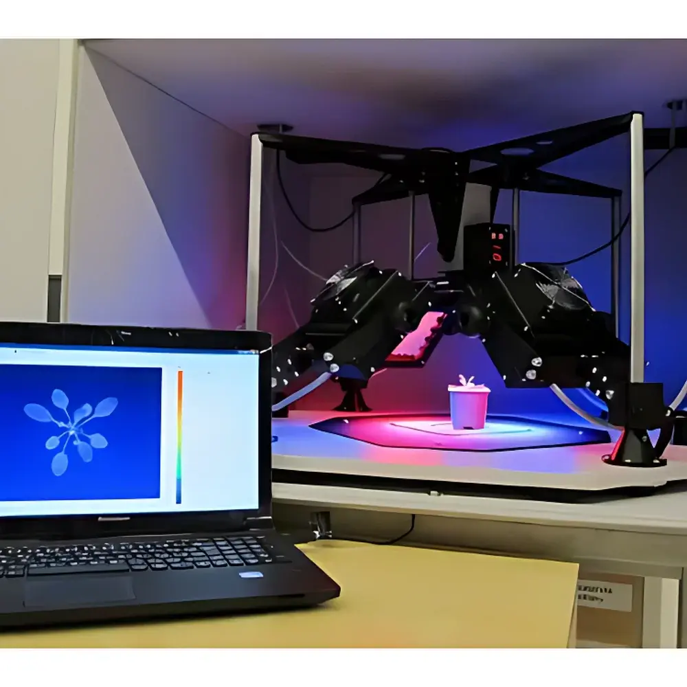
The FluorCam-WIC Modular Plant Phenotyping Imaging System is a high-precision, research-grade platform engineered for non-invasive, quantitative analysis of plant physiological status and stress responses. Based on pulse-amplitude modulated (PAM) chlorophyll fluorescence imaging, multi-spectral UV-induced fluorescence (UV-MCF), and GFP fluorescence protein detection, the system enables spatially resolved, time-series measurements of photosynthetic efficiency, electron transport dynamics, stomatal behavior, metabolic heterogeneity, and thermal regulation across whole plants, detached organs, or high-throughput microplate formats. Designed for controlled-environment laboratories, growth chambers, and phenotyping greenhouses, the FluorCam-WIC adheres to core principles of photobiological measurement integrity—including calibrated photon flux density (PPFD), spectrally defined excitation sources, temporally precise light pulses (10–100 µs), and synchronized CCD acquisition—ensuring reproducibility compliant with ISO 17025-accredited plant physiology workflows.
Key Features
- Modular architecture: Interchangeable imaging modules (PAM, UV-MCF, GFP, IR thermography, RGB) and LED illumination panels enable rapid reconfiguration for diverse experimental objectives.
- Imaging area: 20 × 20 cm standard field-of-view; supports single leaves, floral structures, seedlings (e.g., Arabidopsis), multi-plant arrays, algae cultures, mosses, lichens, and microtiter plates (96-/384-well).
- High-fidelity imaging sensor: TOMI-2 progressive-scan CCD with 1360 × 1024 pixel resolution, 16-bit A/D conversion (65,536 gray levels), 6.45 µm pixel pitch, and up to 20 fps frame rate at full resolution.
- Multi-wavelength illumination: Four independently controllable LED panels (20 × 20 cm active area);标配 617 nm measuring light; dual actinic light (Actinic1: ≤300 µmol·m⁻²·s⁻¹; Actinic2: ≤2000 µmol·m⁻²·s⁻¹; upgradeable to 3000 µmol·m⁻²·s⁻¹); saturating flash up to 4000 µmol·m⁻²·s⁻¹ (upgradeable to 6000).
- Comprehensive parameter quantification: >50 PAM-derived parameters including Fv/Fm, ΦPSII, NPQ, qL, ETR, Rfd, and kinetic traces (Kautsky induction, fluorescence quenching, light response curves).
- Optional spectral expansion: UV-MCF (F440, F520, F690, F740 ratios), PAR absorption & NDVI (735/650 nm LEDs + 7-position filter wheel), GFP/YFP/EGFP detection (470/530 nm excitation + matched emission filters).
- Automated protocol-driven operation: User-defined measurement sequences with programmable timing, light intensities, acquisition intervals, and ROI selection—fully timestamped and stored in hierarchical directory structure.
Sample Compatibility & Compliance
The FluorCam-WIC accommodates live, intact plant material without destructive sampling—ideal for longitudinal studies under controlled abiotic (drought, heat, salinity, UV-B, nutrient deficiency) and biotic (pathogen inoculation, herbivory) stress regimes. It supports compliance with widely adopted plant phenotyping standards, including FAO’s Stress Physiology Protocols, COST Action FA1306 guidelines for chlorophyll fluorescence reporting, and EU-funded transnational access programs (e.g., EMPHASIS, PLANT2020). Data provenance and audit readiness are supported via embedded time-stamping, protocol versioning, and metadata tagging aligned with MIAPPE v1.1 (Minimum Information About a Plant Phenotyping Experiment). While not FDA 21 CFR Part 11-certified out-of-the-box, raw data export (TIFF, CSV, HDF5) and script-based protocol replication facilitate GLP/GMP-aligned validation in regulated breeding or agrochemical efficacy trials.
Software & Data Management
FluorCam software provides a unified interface for real-time monitoring (Live mode), protocol authoring (Protocol Editor with wizard-guided templates and custom scripting), image preprocessing (auto-ROI detection, manual polygonal/sector-based region definition), and quantitative analysis. Two signal-processing modes are implemented: “Signal Calculation then Average” (optimal for high-SNR datasets) and “Signal Average then Calculation” (robust noise suppression for low-light or transient signals). Output includes spatiotemporal fluorescence maps, kinetic curve overlays, statistical summaries per ROI (>1000 regions supported), Excel-exportable parameter tables, histogram distributions, and AVI/MPEG video exports of dynamic fluorescence responses. All acquired images retain EXIF-like metadata (timestamp, light settings, exposure, gain, protocol ID), ensuring traceability for peer-reviewed publication and multi-lab comparative studies.
Applications
- Photosynthetic performance mapping: Spatial heterogeneity of PSII quantum yield, electron transport rate, and photoprotective capacity across leaf lamina or canopy layers.
- Stress phenotyping: Early detection of drought-induced stomatal closure via concurrent chlorophyll fluorescence decline and infrared thermography (WIC module).
- Genetic screening: High-throughput evaluation of mutant or transgenic lines for altered photoprotection, ROS scavenging, or circadian-regulated fluorescence dynamics.
- Pathogen-host interaction: Quantifying localized suppression of photosynthesis preceding visible symptom development (e.g., fungal infection, viral systemic movement).
- Crop breeding support: Correlating fluorescence-derived vigor indices (e.g., Rfd, Fv/Fm) with yield components under field-simulated conditions.
- Morpho-physiological integration: Co-registration of RGB morphology (TetraCam module) with functional fluorescence or thermal signatures for holistic trait modeling.
FAQ
What is the minimum and maximum sample size compatible with the standard FluorCam-WIC configuration?
The system supports samples ranging from individual microplate wells (e.g., 96-well format) to whole rosettes of Arabidopsis or small cereal seedlings within the 20 × 20 cm field of view. Larger specimens require optional macro-lens adaptation or staged scanning protocols.
Can the FluorCam-WIC be integrated into automated growth facilities or robotic phenotyping platforms?
Yes—the system communicates via Gigabit Ethernet and supports external TTL triggering, enabling synchronization with environmental control systems, conveyor-based imaging stations, or robotic arm positioning systems.
Is spectral calibration traceable to NIST standards?
LED peak wavelengths and relative irradiance profiles are factory-characterized using calibrated spectroradiometry; absolute PPFD calibration certificates are available upon request for ISO-compliant validation.
How is data security and long-term archiving handled?
Raw TIFF stacks and processed CSV outputs are stored locally with user-defined folder structures; network-attached storage (NAS) and backup scripts are recommended for institutional data governance compliance.
Does the system support third-party software integration (e.g., Python, MATLAB, R)?
Yes—binary image data (16-bit TIFF) and parameter tables (CSV) are fully interoperable; API documentation and sample scripts for batch processing are provided with the software suite.


