PSI FluorCam Mobile Multispectral Fluorescence Imaging System
| Brand | PSI (Czech Republic) |
|---|---|
| Origin | Czech Republic |
| Model | FluorCam Mobile |
| Imaging Sensor | 1392×1040 CCD, 20 fps, 6.45 µm pixel size |
| Excitation Sources | 4+1 configurable LED array (red, cool white, UV 320–400 nm), optional blue/green/cyan/FAR LEDs |
| Filter Wheel | 7-position motorized, with Chl-F, MCF, GFP/YFP, F440/F520/F690/F740 filters |
| Fluorescence Parameters | >60 quantitative metrics including Fo, Fm, Fv/Fm, NPQ, QY, PARabs, Rfd, BGF, UV-Chl-F ratios |
| NDVI & PAR Absorption | Optional dual-wavelength (630/735 nm) LED module |
| Thermal Imaging (optional) | Uncooled VOx microbolometer, 640×512 pixels, 7.5–13.5 µm, -25 to 150 °C, sensitivity ≤30 mK @ 30 °C |
| RGB Imaging (optional) | 2592×1944 scientific-grade CMOS, 54 dB SNR, 1–40× zoom |
| Power Supply | 90–240 V AC |
| Compliance | CE-marked, ISO/IEC 17025-compatible data acquisition architecture, FDA 21 CFR Part 11-ready software audit trail (with optional configuration) |
Overview
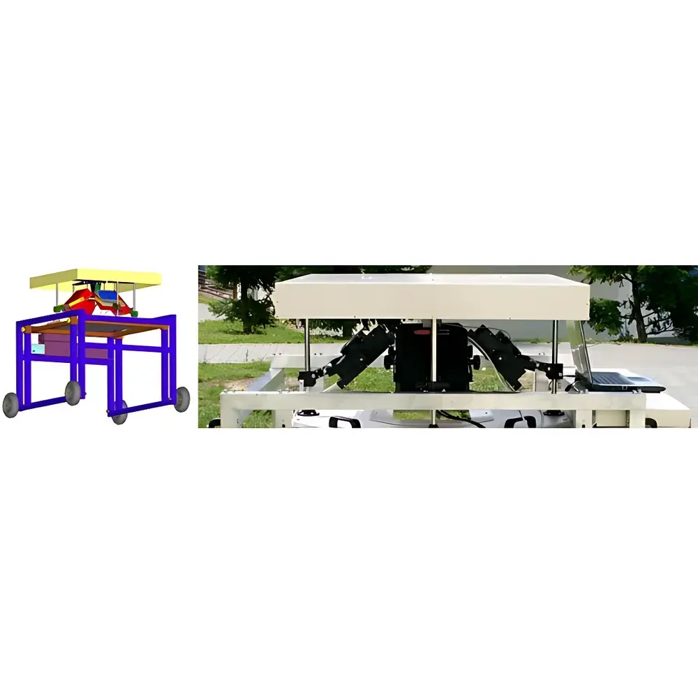
The PSI FluorCam Mobile Multispectral Fluorescence Imaging System is a field-deployable, modular platform engineered for non-invasive, high-resolution physiological phenotyping of plants under controlled or natural environmental conditions. Built upon PSI’s validated pulse-amplitude-modulated (PAM) chlorophyll fluorescence imaging technology, the system extends beyond conventional single-wavelength fluorescence analysis by integrating multispectral excitation and emission detection across ultraviolet (UV), visible, and near-infrared spectral domains. Its core measurement principle relies on quantifying photochemical and non-photochemical energy dissipation pathways in photosystem II (PSII) via kinetic fluorescence induction (Kautsky effect), quenching analysis (NPQ, qP), and spectral fingerprinting of endogenous fluorophores—including chlorophyll *a*, phenolic compounds (F440, F520), and genetically encoded reporters (GFP/YFP). The mobile chassis enables in situ measurements in greenhouses, growth chambers, field plots, or controlled-environment agriculture facilities—eliminating sample transport artifacts while supporting long-term unattended monitoring protocols.
Key Features
- Modular four-wheel mobility platform with adjustable height and terrain-adaptive suspension, certified for indoor and outdoor use (IP65-rated electronics enclosure)
- Configurable 4+1 LED excitation array: dual red (617 nm), dual cool-white (6500 K), and top-mounted UV (320–400 nm) sources—each independently intensity- and pulse-controlled
- High-fidelity 1392×1040 scientific CCD sensor with 20 fps capture rate, 6.45 µm pixel pitch, and hardware binning (2×2 to 4×4) for signal-to-noise optimization
- Motorized 7-position filter wheel supporting simultaneous Chl-F, multispectral UV-induced fluorescence (F440/F520/F690/F740), GFP/YFP, and NDVI-specific bandpass filters
- Programmable dual-protocol automation: two independent experimental sequences (e.g., daytime Kautsky + nighttime NPQ) execute unattended with timestamped data storage
- Integrated spectral calibration workflow compliant with ISO 17025 traceability requirements, enabling inter-laboratory reproducibility
- Optional co-registration modules: uncooled VOx infrared thermal imager (640×512, ±0.03 °C sensitivity) and scientific RGB camera (2592×1944, 54 dB SNR)
Sample Compatibility & Compliance
The FluorCam Mobile accommodates diverse biological specimens without physical contact: detached leaves, intact potted plants, seedlings, fruits, roots (in transparent media), algae suspensions, and small model organisms. Its optical design ensures uniform irradiance over imaging areas of 13×13 cm (standard) or 20×20 cm (large-format option), minimizing edge effects during quantitative spatial mapping. All optical components comply with IEC 62471 photobiological safety standards for UV exposure. Data acquisition adheres to GLP/GMP-aligned metadata tagging—embedding instrument settings, environmental parameters (ambient temperature, humidity, PAR), and operator credentials into each dataset. Software audit trails meet FDA 21 CFR Part 11 requirements when configured with electronic signatures and role-based access control. The system supports ASTM E2912-22 (Standard Guide for Chlorophyll Fluorescence Imaging in Plant Stress Assessment) and ISO 14220-2 (Plant Phenotyping—Imaging Protocols) reference frameworks.
Software & Data Management
FluorCam Acquisition & Analysis Suite v8.x provides a validated, scriptable environment for experimental design, real-time visualization, and statistical modeling. Core modules include Live Preview (for focus/alignment), Protocol Builder (drag-and-drop parameter sequencing), Pre-processing (ROI definition via polygon, ellipse, or freehand tools), and Results Explorer (parametric heatmaps, time-series plots, histogram distributions). Advanced processing modes—“Signal Calculation then Averaging” (for high-SNR datasets) and “Signal Averaging then Calculation” (for low-light conditions)—minimize systematic bias in kinetic parameter derivation. All outputs export to HDF5, TIFF, CSV, or MATLAB-native formats with embedded EXIF metadata. Batch processing pipelines support automated ROI extraction across hundreds of images, while integrated statistical engines compute ANOVA, PCA, and correlation matrices directly within the GUI. Audit logs record every user action, parameter change, and file export event—ensuring full ALCOA+ (Attributable, Legible, Contemporaneous, Original, Accurate) compliance.
Applications
- High-throughput phenotyping for drought, salinity, heavy metal, or pathogen stress screening in breeding programs
- Non-destructive assessment of nitrogen status via NDVI and UV-induced BGF (blue-green fluorescence) ratio dynamics
- Spatiotemporal mapping of PSII photochemical efficiency (ΦPSII), electron transport rate (ETR), and regulatory NPQ components
- Early detection of biotic stressors—including viral infection (e.g., PMMoV), bacterial soft rot (*Dickeya dadantii*), and fungal colonization—through spectral signature divergence
- Functional validation of transgenic lines expressing GFP/YFP reporters under tissue-specific promoters
- Multi-modal integration: correlating chlorophyll fluorescence quenching kinetics with stomatal conductance (via cyan-light-induced response), leaf temperature gradients (IR thermography), and structural traits (RGB morphology)
- Ecophysiological studies of plant-soil-microbe interactions, allelopathy, and pollutant bioindication in field trials
FAQ
What is the minimum required operating distance between the imaging head and sample?
The standard working distance is 50 cm for 13×13 cm illumination area; extended optics support 100 cm for 20×20 cm coverage. Field-of-view scaling maintains optical uniformity per ISO 14220-2 specifications.
Can the system operate autonomously overnight in an unheated greenhouse?
Yes—the chassis and electronics are rated for -15 °C to +50 °C ambient operation. External power must be supplied via industrial-grade UPS or portable generator (≥1.5 kVA, pure sine wave output).
How does the UV excitation module ensure consistent spectral output across repeated measurements?
Each UV LED array undergoes factory spectral calibration against NIST-traceable radiometric standards. Real-time photodiode feedback stabilizes irradiance at ±2% over 8-hour continuous operation.
Is raw data accessible for third-party analysis (e.g., Python or R)?
All image stacks and parameter tables export in open, non-proprietary formats (HDF5, CSV) with complete metadata headers—including excitation wavelengths, filter positions, gain settings, and timestamps.
Does the system support remote monitoring via network connection?
USB 2.0 is standard; optional Gigabit Ethernet interface enables remote control, live streaming, and centralized data aggregation in multi-system deployments.


