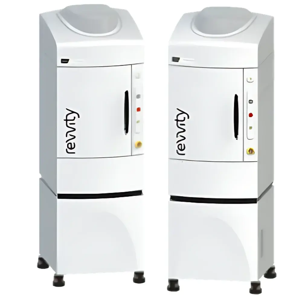
Revvity IVIS Spectrum CT Small Animal In Vivo Optical Imaging System
| Brand | Revvity |
|---|---|
| Origin | USA |
| Manufacturer | Yes |
| Import Status | Imported |
| Model | Spectrum CT |
| Instrument Type | Optical & Micro-CT Hybrid Imaging System |
| Animal Model | Mouse |
| Imaging Modalities | Bioluminescence (2D/3D), Fluorescence (2D/3D), Low-Dose Micro-CT |
| Throughput | Up to 10 mice per scan |
| Camera | eXcelon-coated back-illuminated CCD |
| Software | Living Image® v4.7+ |
| Regulatory Compliance | Designed for GLP-compliant workflows, supports FDA 21 CFR Part 11 audit trail and electronic signature configurations |
Overview
The Revvity IVIS® Spectrum CT is a fully integrated, multimodal preclinical imaging platform engineered for high-sensitivity, quantitative in vivo optical and anatomical imaging in murine models. It combines two complementary modalities—bioluminescence and fluorescence tomography (BLT/FLT) with low-dose, high-resolution micro-computed tomography (micro-CT)—within a single instrument architecture. The system operates on the principle of photon detection via a thermoelectrically cooled, back-illuminated CCD camera featuring proprietary eXcelon coating, which enhances quantum efficiency across the visible-to-near-infrared spectrum (400–900 nm). This enables detection of ultra-low-light signals from luciferase-expressing cells or near-infrared fluorescent probes with exceptional signal-to-noise ratio. Concurrently, the integrated micro-CT module—equipped with a horizontal gantry and flat-panel detector—provides rapid, isotropic volumetric reconstruction (≤50 µm isotropic resolution) with dose optimization algorithms to minimize radiation exposure during longitudinal studies.
Key Features
- eXcelon-coated CCD camera delivering >95% quantum efficiency at 600 nm, enabling sub-picomolar sensitivity for bioluminescent reporter detection
- Simultaneous 2D planar and 3D tomographic reconstruction for both bioluminescence and fluorescence signals, supported by spectral unmixing algorithms to resolve ≥4 spectrally distinct fluorophores
- Integrated micro-CT subsystem with automatic beam hardening correction, iterative reconstruction (OSEM), and dose-efficient acquisition protocols (typical scan time: 3–8 min per mouse at ≤25 mGy)
- High-throughput imaging capability: accommodates up to 10 anesthetized mice in a single session using programmable multi-position staging and intelligent tray alignment
- Precision optical-CT co-registration: hardware-synchronized stage positioning ensures sub-millimeter spatial alignment between functional optical data and anatomical CT volumes
- Modular configuration options: systems may be deployed as standalone IVIS Spectrum 2 (optical-only) or upgraded to IVIS Spectrum CT 2 (hybrid optical + CT) without hardware replacement
Sample Compatibility & Compliance
The IVIS Spectrum CT is validated for use with standard laboratory mouse strains (C57BL/6, BALB/c, nude, NSG) under ISO 14155-compliant study designs. Its optical chamber maintains stable temperature (28–37 °C) and CO2-controlled anesthesia delivery (via integrated vaporizer interface), ensuring physiological consistency across longitudinal sessions. All imaging protocols comply with ARRIVE 2.0 guidelines for reporting animal research. The system meets IEC 61000-6-3 EMC standards and is CE-marked for in vitro diagnostic use where applicable. For regulated environments, Living Image® software supports 21 CFR Part 11 compliance—including role-based access control, electronic signatures, and immutable audit trails—with optional IQ/OQ/PQ documentation packages available.
Software & Data Management
Living Image® software (v4.7 or later) serves as the unified acquisition, processing, and analysis engine. Its wizard-driven interface streamlines protocol setup for BLI, FLI, and CT workflows while preserving full manual control for advanced users. Key capabilities include automated background subtraction, spectral unmixing with reference spectra libraries, 3D surface rendering with CT-guided volume-of-interest (VOI) definition, and kinetic quantification of signal intensity over time. Data export conforms to MIAME/MINRMI standards; raw datasets are stored in vendor-neutral formats (e.g., DICOM for CT, TIFF/OME-TIFF for optical data) compatible with third-party analysis platforms including MATLAB, Python (scikit-image, SimpleITK), and Imaris. Integrated project management tools support metadata tagging, batch processing, and cross-study comparative analytics.
Applications
This platform is routinely deployed in oncology (tumor growth kinetics, metastasis tracking, therapy response monitoring), immunology (cell trafficking, immune checkpoint modulation), infectious disease (pathogen burden quantification), neuroscience (neuroinflammation, gene therapy biodistribution), and drug development (PK/PD correlation, toxicology endpoints). Its ability to quantify molecular signals within anatomically contextualized 3D space makes it particularly valuable for validating target engagement, assessing off-target effects, and supporting IND-enabling nonclinical studies requiring robust, reproducible imaging biomarkers.
FAQ
What is the minimum detectable flux for bioluminescence imaging?
System sensitivity is specified at ≤103 photons/sec/cm2/sr under standardized conditions; actual detection limits depend on exposure time, binning, and biological background.
Can the CT module be used independently of optical imaging?
Yes—the micro-CT subsystem operates autonomously with dedicated acquisition software and can generate DICOM-compliant reconstructions without optical module activation.
Is spectral unmixing limited to specific fluorophores?
No—unmixing is based on user-defined emission spectra; libraries include common dyes (Cy5.5, IRDye800CW, AlexaFluor750) and support custom spectral profiles.
How is anesthesia managed during combined optical-CT sessions?
A single isoflurane delivery line interfaces with both modules via shared manifold; gas flow and concentration are monitored and logged in real time within Living Image.
Does the system support dynamic fluorescence imaging (e.g., FRET or lifetime)?
No—IVIS Spectrum CT is optimized for intensity-based steady-state measurements; time-resolved or lifetime-capable systems require alternative instrumentation.


