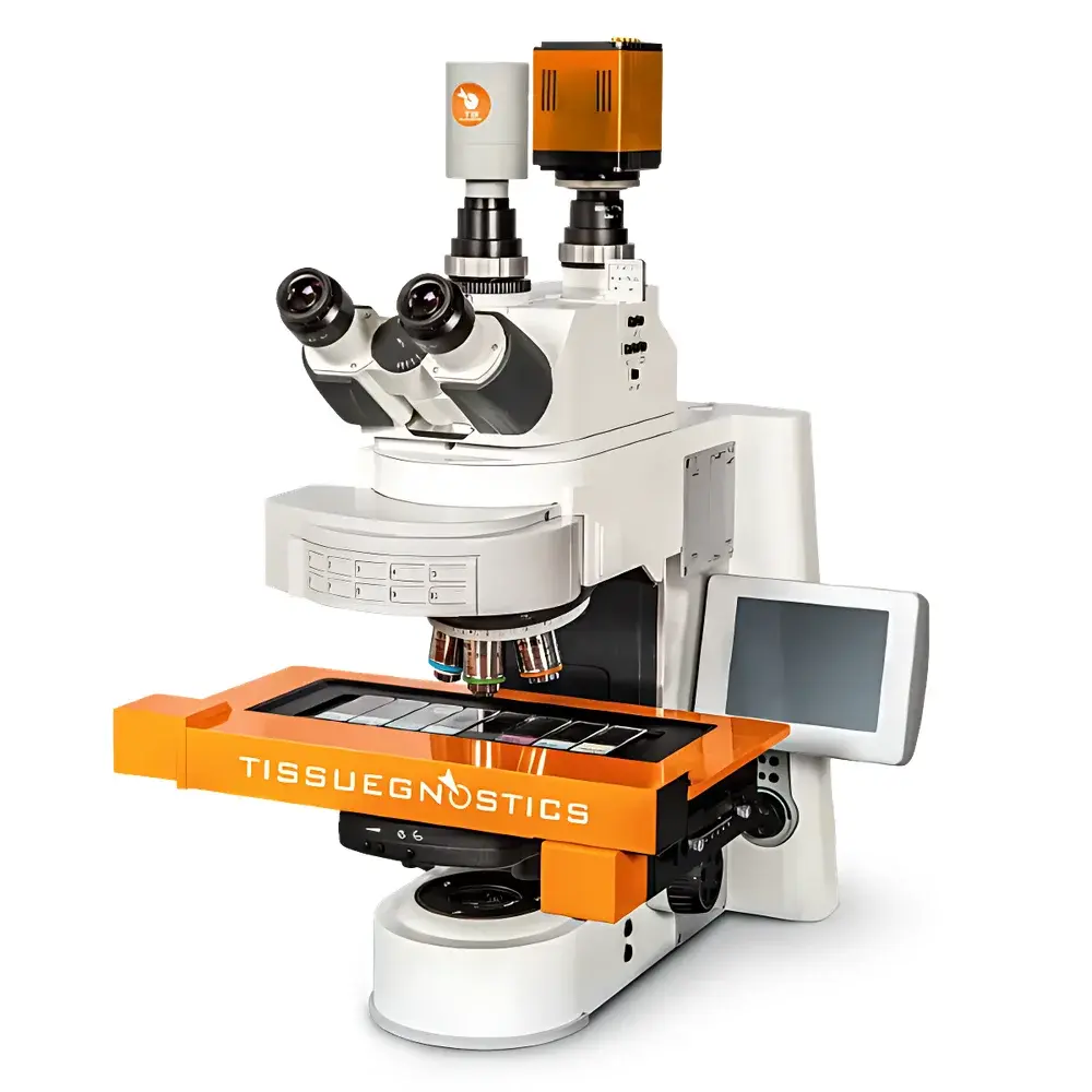
TissueGnostics TISSUEFAXS PLUS In Situ Single-Cell Spatial Phenotyping and Quantification System
| Brand | TissueGnostics |
|---|---|
| Origin | Austria |
| Manufacturer Type | Original Equipment Manufacturer (OEM) |
| Origin Category | Imported Instrument |
| Model | TISSUEFAXS PLUS |
| Price Range | USD 270,000 – 410,000 |
Overview
The TissueGnostics TISSUEFAXS PLUS is a high-content, automated tissue imaging and quantitative analysis platform engineered for in situ single-cell spatial phenotyping. It integrates digital whole-slide imaging with flow cytometry–inspired gating logic to deliver quantitative, location-resolved cellular data directly from intact tissue sections. Unlike conventional image analysis systems that rely solely on pixel-based segmentation or global intensity thresholds, the TISSUEFAXS PLUS applies a dual-modality paradigm: it acquires high-fidelity optical images (brightfield and fluorescence) while simultaneously generating scatterplot-based single-cell event data—each cell annotated with morphometric, intensity, and spatial coordinates (x, y, z). This architecture enables rigorous, reproducible quantification of protein expression, subcellular localization (nucleus/cytoplasm/membrane), and spatial context—including tissue architecture, compartmental distribution (e.g., epithelial vs. stromal), and proximity-based interactions—within native histological context.
Key Features
- Dual independent optical paths for simultaneous brightfield and fluorescence imaging, eliminating cross-channel crosstalk and enabling parallel acquisition of H&E, IHC, IF, and multiplexed immunofluorescence samples.
- Z-stack acquisition support up to 40 focal planes per field, with user-definable step size and adaptive focus refinement using AI-driven, self-evolving autofocus algorithms—optimized for oil-immersion objectives and variable tissue thicknesses.
- Full magnification range compatibility (2.5×–100×), including motorized objective turrets supporting both standard and high-NA oil immersion lenses for subcellular resolution.
- Automated large-format slide handling: supports standard 1×3 inch slides as well as oversized formats (up to 8× scanning factor), with intelligent TMA block recognition and auto-alignment based on fiducial markers and tissue contour detection.
- Integrated channel decomposition for chromogenic brightfield: automatic separation of hematoxylin (nuclear), DAB (membrane/cytoplasmic), and background channels—enabling precise nuclear segmentation and cytomorphometric feature extraction without manual thresholding.
- Context-aware tissue structure recognition: trained classifiers identify epithelial glands, tumor nests, luminal spaces, collagen-rich stroma, and necrotic zones based on texture, shape, and intensity gradients—supporting ISO/IEC 17025-aligned validation workflows.
Sample Compatibility & Compliance
The system processes formalin-fixed paraffin-embedded (FFPE) and frozen tissue sections (4–20 µm), cytospins, and cell pellets. It supports routine IHC (DAB, AEC), multiplex IF (up to 7-color), and H&E-stained slides without modification. All image acquisition and analysis modules comply with FDA 21 CFR Part 11 requirements for electronic records and signatures when deployed with validated software configuration and audit trail logging. Data export formats (TIFF, OME-TIFF, CSV, FCS) conform to MIAME and MIAPE reporting standards. The platform supports GLP/GMP-aligned validation protocols, including instrument qualification (IQ/OQ/PQ), user access control, and version-controlled analysis pipelines traceable to specific slide batches and staining runs.
Software & Data Management
Powered by StrataQuest v8.x, the software provides a unified environment for image acquisition, batch processing, and statistical analysis. It implements hierarchical gating (Input Gate → Primary Gate → Secondary Gate) analogous to flow cytometry, allowing sequential filtering of cells by size, intensity, shape, and spatial constraints (e.g., “CD3+ T cells within 50 µm of PD-L1+ macrophages”). All gates are bi-directionally linked: selecting a population in a scatterplot instantly highlights corresponding cells in the registered tissue image—and vice versa. Audit trails record every parameter change, gate adjustment, and export action with timestamp, user ID, and reason code. Data exports include spatial heatmaps, neighborhood interaction matrices, and FCS files compatible with FlowJo and Cytobank for cross-platform validation.
Applications
- Tumor microenvironment characterization: quantifying immune infiltrate density, spatial segregation of checkpoint ligands/receptors, and tertiary lymphoid structure (TLS) mapping.
- Translational biomarker development: correlating protein co-expression patterns (e.g., pERK + Ki67) with histopathological grade and clinical outcome in retrospective cohorts.
- Human Proteome Project integration: cross-referencing spatial protein abundance data against the Human Protein Atlas annotation framework for tissue-specific expression validation.
- Stem cell niche analysis: identifying rare undifferentiated progenitors via combinatorial marker gating (e.g., SOX2+/CD44−/Ki67low) and validating their anatomical positioning relative to vascular or stromal cues.
- Preclinical toxicology: detecting subtle morphometric shifts in hepatocyte nuclei or renal tubular epithelium following compound exposure—quantified across hundreds of fields with statistical confidence intervals.
FAQ
Does TISSUEFAXS PLUS support multiplexed fluorescence imaging with spectral unmixing?
Yes—when configured with a tunable filter-based or hyperspectral camera module, the system performs linear unmixing of up to 7 fluorophores using reference spectra acquired from single-stain controls.
Can analysis pipelines be locked and validated for regulated environments?
Yes—StrataQuest supports SOP-mode operation with password-protected pipeline templates, immutable parameter sets, and full audit trail generation compliant with 21 CFR Part 11 Annex 11 and ISO 13485.
Is Z-stack data used for 3D reconstruction or only for extended depth-of-field (EDF) synthesis?
Both: EDF synthesis is applied for rapid screening; raw Z-stack volumes are retained for volumetric morphometry (e.g., nuclear sphericity, organelle distribution) and colocalization analysis in 3D space.
How does the system handle tissue folding or section wrinkling during analysis?
The software includes warp-correction algorithms that register each field using tissue-edge contours and landmark-based elastic alignment—preserving spatial fidelity without requiring manual intervention.
Can results be exported to LIMS or enterprise data warehouses?
Yes—via RESTful API and configurable ODBC/JDBC connectors, supporting direct ingestion into LabVantage, Thermo Fisher SampleManager, or custom SQL-based repositories with metadata schema mapping.


