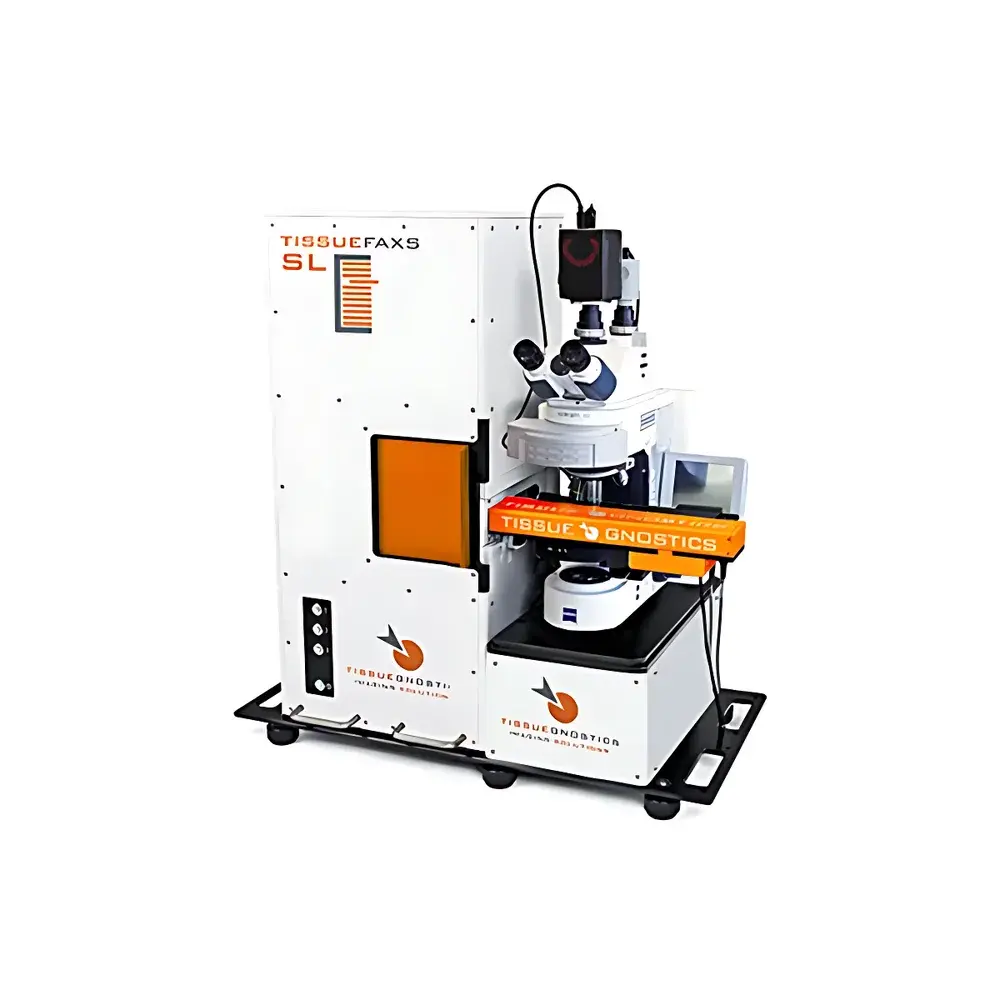
TissueGnostics TissueFAXS SL Slide Loader System
| Brand | TissueGnostics |
|---|---|
| Country of Origin | Austria |
| Manufacturer Type | Original Equipment Manufacturer (OEM) |
| Import Status | Imported Instrument |
| Model | TISSUEFAXS SL SLIDE LOADER |
| Price Range | USD 420,000 – 700,000 |
Overview
The TissueGnostics TissueFAXS SL Slide Loader System is a fully automated, high-throughput whole-slide imaging (WSI) platform engineered for quantitative tissue and cell analysis in translational research and digital pathology laboratories. Built upon the proven TissueFAXS optical architecture—compatible with upright configurations including Fluo, Histo, Plus, Spectra, and Q+ variants—the SL variant integrates a robotic slide loader, precision motion control, and real-time image quality assurance to enable unattended, reproducible acquisition of brightfield, fluorescence, and tissue microarray (TMA) specimens. Its core imaging principle relies on motorized stage scanning combined with high-resolution objective lenses (2.5× to 100×), synchronized with scientific-grade CMOS or sCMOS cameras, to generate gigapixel-scale virtual slides compliant with DICOM-SR and OpenSlide standards. Designed for GLP- and GCP-aligned workflows, the system supports traceable acquisition metadata, audit-ready logging, and integration into LIMS and PACS environments.
Key Features
- Automated slide handling capacity: Up to 120 standard glass slides (76 × 26 mm) per run via three 40-slot slide cassettes—each cassette holds 20 dual-position carriers (2 slides per carrier)
- Alternative loading mode: Supports up to 60 extended-format slides (152 × 26 mm) using dedicated cassettes
- Non-contact magnetic gripper mechanism: Eliminates mechanical pressure or abrasion on tissue sections during loading/unloading, preserving sample integrity and minimizing section delamination risk
- Real-time focus validation (SV – Slide Validator): Analyzes sharpness metrics across every field-of-view (FOV) during acquisition; automatically re-scans out-of-focus regions without user intervention
- Vibration-damped optical bench: Integrated passive isolation platform minimizes stage drift and optical misalignment during long-duration scans
- Multi-modal compatibility: Native support for brightfield (H&E, IHC), fluorescence (DAPI, FITC, TRITC, Cy5), and multi-spectral imaging when paired with TissueFAXS Spectra hardware
- Objective flexibility: Full compatibility with 2.5×, 5×, 10×, 20×, 40×, and 100× Plan-Apochromat objectives—including oil-immersion and water-dipping variants—for scalable resolution from overview mapping to subcellular detail
Sample Compatibility & Compliance
The TissueFAXS SL accommodates standard histological glass slides (1–1.2 mm thickness), charged or poly-L-lysine-coated substrates, and specialty formats including cytology smears, frozen sections, and decalcified bone preparations. It maintains consistent Z-stack acquisition across variable tissue thicknesses and mounting media refractive indices. All hardware and software components comply with CE marking requirements under Directive 2014/30/EU (EMC) and 2014/35/EU (LVD). The system supports 21 CFR Part 11-compliant electronic signatures and audit trails when deployed with validated TissueFAXS software version 5.0 or later. Data export formats include TIFF (uncompressed), OME-TIFF, NDPI-compatible pyramidal tiff, and DICOM WSI objects—ensuring interoperability with Qupath, HALO, Visiopharm, and Aperio ImageScope platforms.
Software & Data Management
TissueFAXS Software v5.x provides two operational modes: Simple Mode for homogeneous slide batches (e.g., routine IHC screening) and Advanced Mode for heterogeneous cohorts requiring per-slide parameter overrides (e.g., mixed fluorophore panels, variable exposure times, or multi-channel registration). The embedded Slide Validator (SV) module performs real-time MTF-based sharpness scoring at each FOV using normalized gradient magnitude variance. Acquisition logs record timestamp, objective ID, exposure settings, stage coordinates, focus offset, and SV confidence score per tile. Raw data are stored in hierarchical folder structures with SHA-256 checksums; processed virtual slides support metadata embedding per ISO 13606 and HL7 FHIR ImagingStudy resources. Optional TissueMINER integration enables on-the-fly AI-assisted segmentation and biomarker quantification directly within the acquisition pipeline.
Applications
- Quantitative immunohistochemistry (qIHC) and multiplexed IF (mIF) analysis in oncology biomarker studies (e.g., PD-L1, CD8, Ki67 spatial profiling)
- TMA construction and high-content screening across hundreds of patient-derived samples
- Neuropathology applications including amyloid plaque load quantification and neuronal morphology mapping
- Toxicologic pathology assessments requiring standardized cross-study comparison of lesion burden and distribution
- Preclinical drug development workflows where longitudinal tissue response must be tracked across large animal cohorts
- Digital pathology training set generation for deep learning model development under HIPAA- and GDPR-compliant infrastructure
FAQ
Does the TissueFAXS SL support autofocus during scanning?
Yes—autofocus is performed at every FOV using contrast-based algorithms optimized for both brightfield and fluorescence contrast; optional hardware-based laser triangulation focus sensors are available for ultra-thick sections.
Can the system integrate with existing LIS/PACS infrastructure?
Yes—via DICOM WSI conformance (Supplement 145), HL7 ADT messaging, and configurable RESTful API endpoints for bidirectional data exchange.
What slide barcode standards are supported?
GS1-128, Code 128, QR Code, and DataMatrix ECC200; custom OCR-based label recognition modules can be deployed for legacy handwritten identifiers.
Is remote monitoring and control possible?
Yes—through TissueFAXS Remote Access Server (TRAS), which provides encrypted web-based status dashboards, live scan progress tracking, and emergency pause/resume functionality.
How is calibration traceability maintained?
Each instrument ships with NIST-traceable stage calibration certificates; periodic verification is supported using ISO 10993-qualified calibration slides with certified feature spacing and intensity gradients.
Related Products


